

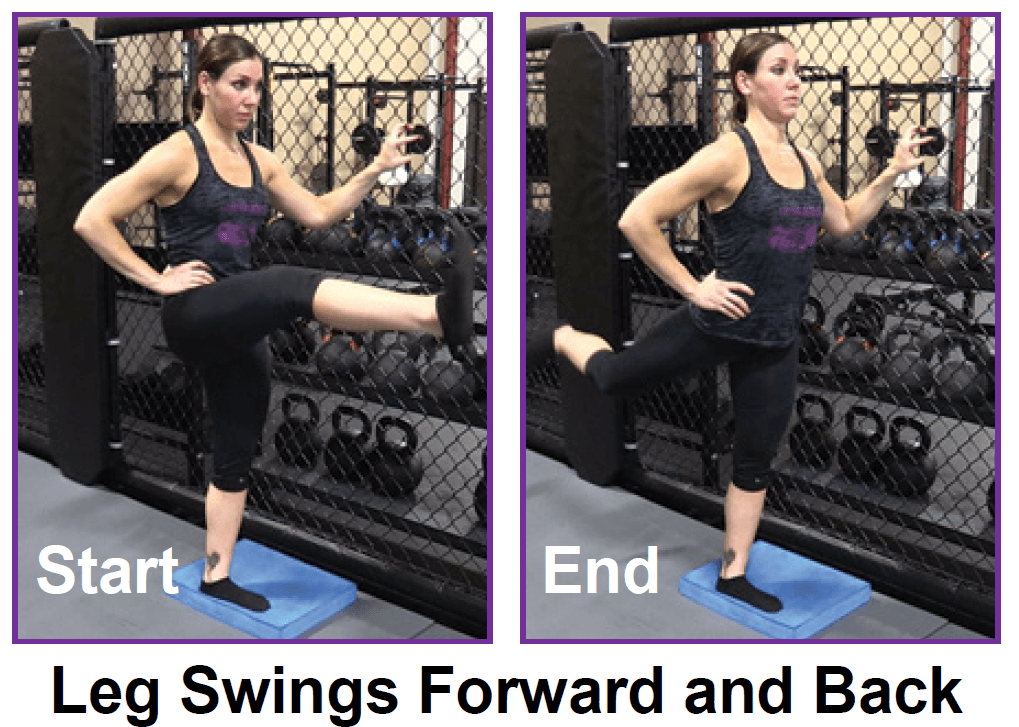
To help you strengthen these important muscles, Miranda put together a list of exercises, below. They include dynamic warm-up moves, meant to activate your hip muscles and prep them for the bigger movements to come; functional moves that train basic movement patterns, like the squat, hip hinge, and lunge; functional plyometric exercises that train explosive power; and a few moves that get you moving in different planes of motion, or directions.
The hip is a common site of osteoarthritis. To help protect the hip joint from "wear and tear," it is important to strengthen the muscles that support it. Your hip also controls the position of your knee, and strengthening your hips may be one component of your rehab program for knee pain. Your physical therapist may also prescribe hip exercises after total hip replacement if you have a hip labrum tear or as part of your hip exercise program for hip pain.
People understandably assume that the worst back pain is the scariest. In fact, pain intensity is a poor indicator of back pain ominousness,10 and some of the worst causes are actually the least painful (especially in the early stages). For instance, someone could experience the symptoms of cauda equinae syndrome, and be in real danger of a serious and permanent injury to their spine, but have surprisingly little pain — even none at all in some cases!
This information is not designed to replace a physician's independent judgment about the appropriateness or risks of a procedure for a given patient. Always consult your doctor about your medical conditions or back problem. SpineUniverse does not provide medical advice, diagnosis or treatment. Use of the SpineUniverse.com site is conditional upon your acceptance of our User Agreement
How to do it: Stand with your feet together, holding dumbbells by your sides. Take a wide step out to your right and lower into a side lunge, reaching dumbbells on either side of right leg [as shown]. Bend your left knee and shift your weight into both legs, into a wide squat position, reaching the dumbbells to floor in front of you, then extend your right leg and shift your weight to the left, moving into a side lunge with your left leg.
The bony lumbar spine is designed so that vertebrae "stacked" together can provide a movable support structure while also protecting the spinal cord from injury. The spinal cord is composed of nervous tissue that extends down the spinal column from the brain. Each vertebra has a spinous process, a bony prominence behind the spinal cord, which shields the cord's nervous tissue from impact trauma. Vertebrae also have a strong bony "body" (vertebral body) in front of the spinal cord to provide a platform suitable for weight bearing of all tissues above the buttocks. The lumbar vertebrae stack immediately atop the sacrum bone that is situated in between the buttocks. On each side, the sacrum meets the iliac bone of the pelvis to form the sacroiliac joints of the buttocks.
If low back pain occurs after a recent injury — such as a car accident, a fall or sports injury — call your primary-care physician immediately. If there are any neurological symptoms, seek medical care immediately. If there are no neurological problems (i.e. numbness, weakness, bowel and bladder dysfunction), the patient may benefit by beginning conservative treatment at home for two to three days. The patient may take anti-inflammatory medications such as aspirin or ibuprofen and restrict strenuous activities for a few days.
How to: Sit down with your knees bent and feet flat on the floor in front of you (a). Place your right ankle on top of your left thigh and flex your right foot (b). Put your hands behind your body, fingertips facing away from your body and begin to press your hips toward your heels until you feel a stretch through your outer left hip. Keep your back tall and chest open (c). Hold for six to eight breaths, then repeat on the other side.
Vertebroplasty and kyphoplasty are minimally invasive treatments to repair compression fractures of the vertebrae caused by osteoporosis. Vertebroplasty uses three-dimensional imaging to assist in guiding a fine needle through the skin into the vertebral body, the largest part of the vertebrae. A glue-like bone cement is then injected into the vertebral body space, which quickly hardens to stabilize and strengthen the bone and provide pain relief. In kyphoplasty, prior to injecting the bone cement, a special balloon is inserted and gently inflated to restore height to the vertebral structure and reduce spinal deformity.
How to: Stand tall, feet slightly wider than shoulder-width apart, toes pointed out at 45-degree angles (a). Keep your back straight, knees over toes and your weight in the heels of your feet (b). Engage your glutes and thighs as you lower into a deep squat until thighs parallel to the ground (or as close as you can get them) (c). Powering through your heels, push up to return to starting position (d). Repeat.

Start in a runner’s lunge with right leg forward, right knee over right ankle and back leg straight. Walk right foot over toward left hand, then drop right shin and thigh to the floor, making sure to keep right knee in line with right hip. Allow left leg to rest on the floor with top of left foot facing down. Take a moment to square your hips to the front of the room. Hold here, or hinge at hips and lower torso toward floor, allowing head to rest on forearms. Hold for at least 30 seconds, then repeat on opposite side. You want to feel a moderate stretch in the outside of the right thigh, but if this pose hurts your knees or feels too uncomfortable, stick with Thread the Needle.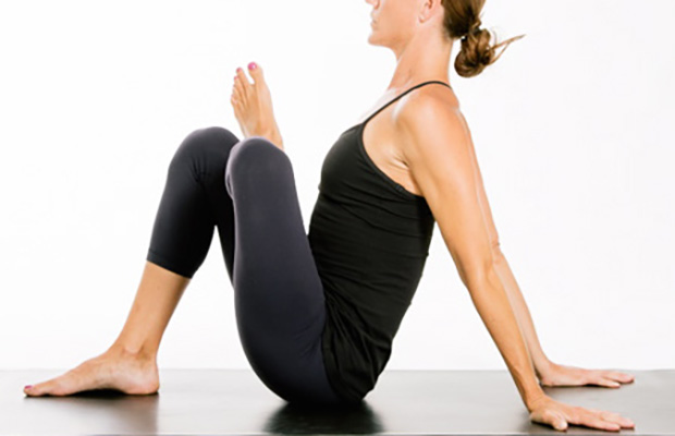
The hip flexors are the group of muscles that allow you to lift your knees toward your chest and bend forward from the hips. What is collectively referred to as the hip flexors is actually a group of muscles that includes the iliopsoas, the thigh muscles (rectus femoris, Sartorius and tensor fasciae latae), and the inner thigh muscles (adductor longus and brevis, pectineus and gracilis).
Chronic back pain is defined as pain that persists for 12 weeks or longer, even after an initial injury or underlying cause of acute low back pain has been treated. About 20 percent of people affected by acute low back pain develop chronic low back pain with persistent symptoms at one year. In some cases, treatment successfully relieves chronic low back pain, but in other cases pain persists despite medical and surgical treatment.
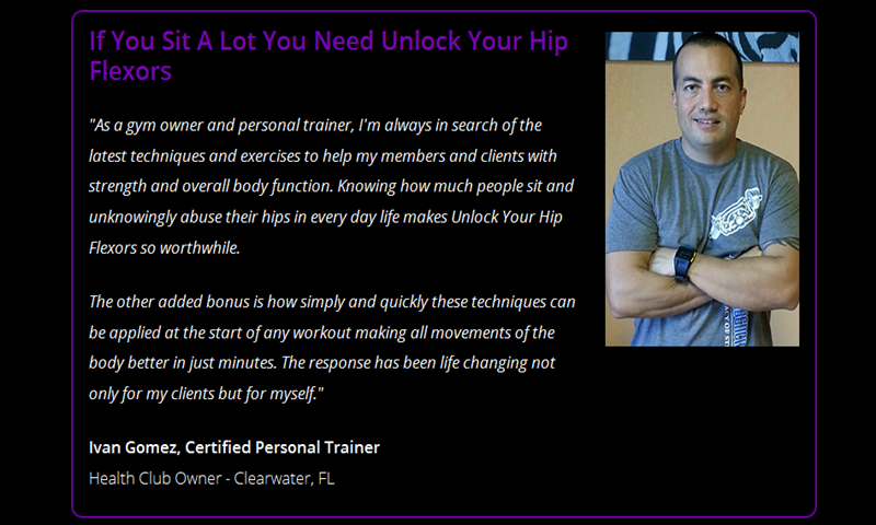
Luckily, you don’t have to quit your day job or forgo spin class to loosen them up. Simply stretching those hips can get your body back in alignment, increase your mobility (and thus your exercise performance) and maybe even ease pesky back pain, Moore says. “Given the amount of time we sit [each] day and the stress we put our bodies under, hip-opening moves are a necessary party of our daily routine.”
Pain on the outside of the hip is most commonly due to greater trochanteric bursitis. The greater trochanter is the protrusion where the thigh bone juts outward at the base of the neck (which connects the ball to the femur and is the site of hip stress fractures). A lubricating sac (or bursa) lies over the boney protrusion so that the surrounding muscles do not rub directly on the bone. The top region of the iliotibial (IT) band, known as the tensor fascia lata, is commonly involved in greater trochanteric bursitis.
First and foremost, stop slouching. One of the most common causes of low back pain is poor sitting posture. The strain on the back while sitting in a slouched position can cause excessive pressure on the joints, muscles, and discs, causing pain. Learn to sit with correct posture and maintain that posture at all times to help decrease or eliminate your low back pain. Also be sure your workspace is set up properly at home and at work.

Increasing general physical activity has been recommended, but no clear relationship to pain or disability has been found when used for the treatment of an acute episode of pain.[48][54] For acute pain, low- to moderate-quality evidence supports walking.[55] Treatment according to McKenzie method is somewhat effective for recurrent acute low back pain, but its benefit in the short term does not appear significant.[1] There is tentative evidence to support the use of heat therapy for acute and sub-chronic low back pain[56] but little evidence for the use of either heat or cold therapy in chronic pain.[57] Weak evidence suggests that back belts might decrease the number of missed workdays, but there is nothing to suggest that they will help with the pain.[50] Ultrasound and shock wave therapies do not appear effective and therefore are not recommended.[58][59] Lumbar traction lacks effectiveness as an intervention for radicular low back pain.[60]
Results after four years of follow-up showed that in general, otherwise healthy people who have surgery for one of these three conditions are likely to fare better than those who receive non-operative care. However, the results also indicated that people who are reluctant to have surgery may also recover with non-operative treatments if their conditions are not progressing and their pain is tolerable, and importantly, delaying or avoiding surgery did not cause additional damage in most cases. Researchers are continuing to track SPORT patient cohorts over a nine-year follow-up period to assess longer term treatment results and cost effectiveness across treatment options. In the interest of improving surgical techniques, NIH also is funding research on factors that contribute to the success or failure of artificial disc replacement surgery, including studies to compare discs on the market for significant differences in their durability rates over time.
Epidural injections of steroid drugs are frequently used to treat sciatica, despite limited evidence for their effectiveness. Moreover, these treatments are based on the assumption that reducing local inflammation in the vertebral column will relieve pain, but an association between structural abnormalities, inflammation, and sciatica symptoms has not been clearly demonstrated. NINDS-funded researchers are using a new imaging technique that can detect inflammation to better understand what causes chronic sciatica pain and to provide evidence to inform treatment selection.

If you have arthritis or bursitis, you’ve probably noticed that exercise can actually help relieve your pain. But when is hip pain a sign that you should stop exercising or doing a certain activity? If your hip starts to hurt during a particular exercise and lingers for hours or days afterward, that’s a sign that your joint needs to rest, notes Humphrey. It’s normal to feel some soreness the day after exercising, but the pain shouldn’t persist or become worse. Also, if you experience a sharp or shooting pain, stop the activity immediately and talk to your doctor or physical therapist.
Doing the bridge exercise in the morning gets your muscles working, activated, and engaged and will help support you the rest of the day, says Humphrey. Lie on your back with your legs bent and your feet flat on the floor, hip-width apart. Press down through your ankles and raise your buttocks off the floor while you tighten your abdominal muscles. Keep your knees aligned with your ankles and aim for a straight line from knees to shoulders, being sure not to arch your back; hold this position for three to five seconds and then slowly lower your buttocks back to the floor. Start with one set of 10 and build up to two or three sets.

Their research differs from past studies of chronic low back pain, which tended to focus on patients who already had a well-established track record of long-term problems (in other words, the people who had already drawn the short straw before they were selected for study, and are likely to carry right on feeling rotten). Instead they studied new cases of chronic low back pain, and found that “more than one third” recovered within nine more months. This evidence is a great foundation for more substantive and lasting reassurance for low back pain patients.
Magnetic resonance imaging (MRI) uses a magnetic force instead of radiation to create a computer-generated image. Unlike x-ray, which shows only bony structures, MRI scans also produce images of soft tissues such as muscles, ligaments, tendons, and blood vessels. An MRI may be ordered if a problem such as infection, tumor, inflammation, disc herniation or rupture, or pressure on a nerve is suspected. MRI is a noninvasive way to identify a condition requiring prompt surgical treatment. However, in most instances, unless there are “red flags” in the history or physical exam, an MRI scan is not necessary during the early phases of low back pain.
Acupuncture is moderately effective for chronic low back pain. It involves the insertion of thin needles into precise points throughout the body. Some practitioners believe this process helps clear away blockages in the body’s life force known as Qi (pronounced chee). Others who may not believe in the concept of Qi theorize that when the needles are inserted and then stimulated (by twisting or passing a low-voltage electrical current through them) naturally occurring painkilling chemicals such as endorphins, serotonin, and acetylcholine are released. Evidence of acupuncture’s benefit for acute low back pain is conflicting and clinical studies continue to investigate its benefits.
Contour Sleep Knee Spacer: Correct sleep alignment is a critical component to rehabilitating an injured hip. This device can help to decrease pressure to the legs and hips while you sleep. Perfect for side sleepers, the Contour Sleep Knee Spacer fits softly between the knees without disrupting your sleep. It helps tense muscles relax and lets you have a better night’s sleep free from painful hip tension.
Most functional exercises—ones that mimic everyday movements such as squats, hip hinges (deadlifts, for example), lunges, steps-ups—stretch and strengthen your hip muscles in some way. So if you strength train and do a variety of these sorts of movements, you're probably working these important muscles without even realizing it. On the other hand, if you mostly focus on exercise methods that have you doing the same movement over and over again, like running or cycling, there's a good chance your hips aren't as strong as they should be. And that can have a negative impact on not only your workouts, but how you move through life in general.
Shingles (herpes zoster) is an acute infection of the nerves that supply sensation to the skin, generally at one or several spinal levels and on one side of the body (right or left). Patients with shingles usually have had chickenpox earlier in life. The herpes virus that causes chickenpox is believed to exist in a dormant state within the spinal nerve roots long after the chickenpox resolves. In people with shingles, this virus reactivates to cause infection along the sensory nerve, leading to nerve pain and usually an outbreak of shingles (tiny blisters on the same side of the body and at the same nerve level). The back pain in patients with shingles of the lumbar area can precede the skin rash by days. Successive crops of tiny blisters can appear for several days and clear with crusty inflammation in one to two weeks. Patients occasionally are left with a more chronic nerve pain (postherpetic neuralgia). Treatment can involve symptomatic relief with lotions, such as calamine, or medications, such as acyclovir (Zovirax), for the infection and pregabalin (Lyrica) or lidocaine (Lidoderm) patches for the pain.
Sciatica is a form of radiculopathy caused by compression of the sciatic nerve, the large nerve that travels through the buttocks and extends down the back of the leg. This compression causes shock-like or burning low back pain combined with pain through the buttocks and down one leg, occasionally reaching the foot. In the most extreme cases, when the nerve is pinched between the disc and the adjacent bone, the symptoms may involve not only pain, but numbness and muscle weakness in the leg because of interrupted nerve signaling. The condition may also be caused by a tumor or cyst that presses on the sciatic nerve or its roots.
Sciatica is a form of radiculopathy caused by compression of the sciatic nerve, the large nerve that travels through the buttocks and extends down the back of the leg. This compression causes shock-like or burning low back pain combined with pain through the buttocks and down one leg, occasionally reaching the foot. In the most extreme cases, when the nerve is pinched between the disc and the adjacent bone, the symptoms may involve not only pain, but numbness and muscle weakness in the leg because of interrupted nerve signaling. The condition may also be caused by a tumor or cyst that presses on the sciatic nerve or its roots.
For persistent low back pain, the short-term outcome is also positive, with improvement in the first six weeks but very little improvement after that. At one year, those with chronic low back pain usually continue to have moderate pain and disability.[2] People at higher risk of long-term disability include those with poor coping skills or with fear of activity (2.5 times more likely to have poor outcomes at one year),[96] those with a poor ability to cope with pain, functional impairments, poor general health, or a significant psychiatric or psychological component to the pain (Waddell's signs).[96]
MRI and x-ray for low back pain are surprisingly unreliable,1 because things like bulging discs usually aren’t a deal,2 most back pain goes away on its own,3 and trigger points (“muscle knots”) are common and can be alarmingly intense but aren’t dangerous.4 Most patients are much better off when they feel confident about these things. The power of justified, rational confidence is a huge factor in back pain.5 Sadly, many healthcare professionals continue to perpetuate the idea of fragile backs,6 which undermines that valuable confidence.

Hamstring squeeze. Use the machine that works your hamstrings; you will either lie on your stomach or sit with a pad behind your knee. Push against the pad, moving your knee up toward the ceiling or backward (depending on which position you’re in). “In other words, bend your knees,” Pariser says. But to avoid cramps in your hamstring muscles, don’t bend your knee so much that your heels are too close to your buttocks. 
Talmage, J; Belcourt, R; Galper, J; et al. (2011). "Low back disorders". In Kurt T. Hegmann. Occupational medicine practice guidelines : evaluation and management of common health problems and functional recovery in workers (3rd ed.). Elk Grove Village, IL: American College of Occupational and Environmental Medicine. pp. 336, 373, 376–377. ISBN 978-0615452272.
Cat-cow: Start on all fours, with palms and knees on the ground. Slowly arch your back, tilt your butt up and look up at the ceiling so you make a C curve with your torso. Return to the neutral starting position, then tilt your pelvis down and gaze at the floor near your feet, so your body reverses the C curve the other way. Return to neutral and repeat the series three times.
To help you strengthen these important muscles, Miranda put together a list of exercises, below. They include dynamic warm-up moves, meant to activate your hip muscles and prep them for the bigger movements to come; functional moves that train basic movement patterns, like the squat, hip hinge, and lunge; functional plyometric exercises that train explosive power; and a few moves that get you moving in different planes of motion, or directions.
Stretching is your next move, but not just any stretches. “Before your workout, you want to go for dynamic stretches, or stretches that put the joint through a full range of motion,” says Lefkowith. Moves like squats and lunges will get your muscles fired up (especially if you focus on squeezing your butt at the top of those squats), says Lefkowith.
Iliopsoas syndrome, which is also called psoas syndrome or iliopsoas tendonitis, occurs when the iliopsoas muscles are injured. Lower back pain is the most common symptom; however, pain can also occur in the hip, thigh, or leg. The iliopsoas bursa, which is a fluid-filled sac located on the inside of the hip that reduces rubbing and friction, is also likely to become inflamed due to the proximity of the two structures. When this happens, the inflamed bursae will make it difficult to move.

