Arthritis: The spondyloarthropathies are inflammatory types of arthritis that can affect the lower back and sacroiliac joints. Examples of spondyloarthropathies include reactive arthritis (Reiter's disease), ankylosing spondylitis, psoriatic arthritis, and the arthritis of inflammatory bowel disease. Each of these diseases can lead to low back pain and stiffness, which is typically worse in the morning. These conditions usually begin in the second and third decades of life. They are treated with medications directed toward decreasing the inflammation. Newer biologic medications have been greatly successful in both quieting the disease and stopping its progression.
But moving is important for hip and knee OA. It causes your joints to compress and release, bringing blood flow, nutrients, and oxygen into the cartilage. “This can help prolong the function and longevity of your joints,” says Eric Robertson, DPT, a physical therapist and associate professor of clinical physical therapy at the University of Southern California. 
Dr. Shiel received a Bachelor of Science degree with honors from the University of Notre Dame. There he was involved in research in radiation biology and received the Huisking Scholarship. After graduating from St. Louis University School of Medicine, he completed his Internal Medicine residency and Rheumatology fellowship at the University of California, Irvine. He is board-certified in Internal Medicine and Rheumatology.
These are really great tips. Just to imform my friends here, my cousin also gave me this link about some other techniches you can use. You have to know exactly what is going on in your body you know. the product is called Panifix, or "Unlock your hip flexor" which Gives You A Practical, Easy-to-follow Program You Can Use To Instantly Release Your Hip Flexors For More Strength, Better Health And All Day Energy. Proven Swipes And Creatives Here:https://tinyurl.com/yd6nbzfh 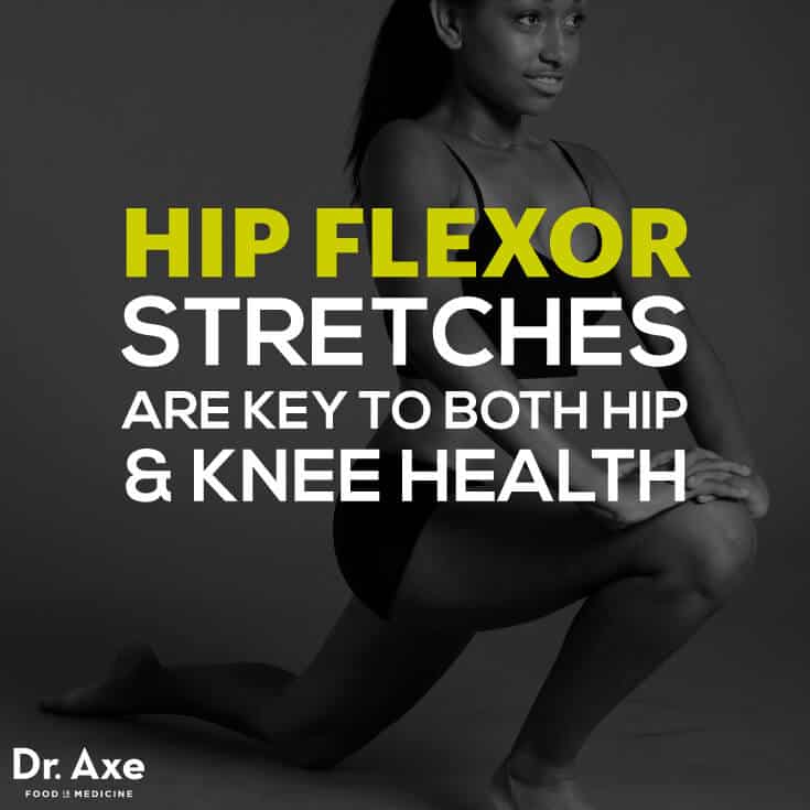
Veritas Health publishes original and accessible health related content written by more than 100 physician authors and peer-reviewed by a 16 member Medical Advisory Board. The Veritas Health platform comprising of Spine-health.com, Arthritis-health.com, Sports-health.com, and Pain-health.com, provides comprehensive information on back pain, arthritis, sports injuries, and chronic pain conditions. For more information visit Veritashealth.com.
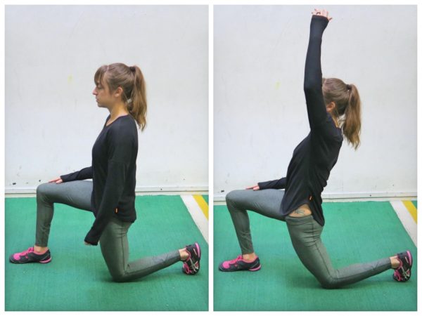
Apply the above concept to your hips. When you sit, your hips are in a "flexed" position. Therefore, the muscles that flex your hips are in a shortened state. You probably spend at least a third of your day sitting down. Think about how much time those hip flexor muscles stay shortened. A lot. Over time, they become tighter and tighter until you look like the old man in the picture. So unless you want to look like that, perform the stretches shown below.
Electrodiagnostics are procedures that, in the setting of low back pain, are primarily used to confirm whether a person has lumbar radiculopathy. The procedures include electromyography (EMG), nerve conduction studies (NCS), and evoked potential (EP) studies. EMG assesses the electrical activity in a muscle and can detect if muscle weakness results from a problem with the nerves that control the muscles. Very fine needles are inserted in muscles to measure electrical activity transmitted from the brain or spinal cord to a particular area of the body. NCSs are often performed along with EMG to exclude conditions that can mimic radiculopathy. In NCSs, two sets of electrodes are placed on the skin over the muscles. The first set provides a mild shock to stimulate the nerve that runs to a particular muscle. The second set records the nerve’s electrical signals, and from this information nerve damage that slows conduction of the nerve signal can be detected. EP tests also involve two sets of electrodes—one set to stimulate a sensory nerve, and the other placed on the scalp to record the speed of nerve signal transmissions to the brain.
I am a science writer, former massage therapist, and I was the assistant editor at ScienceBasedMedicine.org for several years. I have had my share of injuries and pain challenges as a runner and ultimate player. My wife and I live in downtown Vancouver, Canada. See my full bio and qualifications, or my blog, Writerly. You might run into me on Facebook or Twitter.
Medicine FinderLatest NewsSlip, slap, slop early to save your skinComplementary medicine users have worse cancer survivalVideo: BoredomPut down the tablet; pick up a bookVideo: Lift your mood with some weightsQuicken your step to live longerThis web site is intended for Australian residents and is not a substitute for independent professional advice. Information and interactions contained in this Web site are for information purposes only and are not intended to be used to diagnose, treat, cure or prevent any disease. Further, the accuracy, currency and completeness of the information available on this Web site cannot be guaranteed. Tonic Digital Media Pty Ltd, its affiliates and their respective servants and agents do not accept any liability for any injury, loss or damage incurred by use of or reliance on the information made available via or through myDr whether arising from negligence or otherwise. See Privacy Policy and Disclaimer.2001-2019 myDr.com.au © | All Rights Reserved About UsContact UsDisclaimerPrivacy PolicyAdvertising PolicySitemap

Veritas Health publishes original and accessible health related content written by more than 100 physician authors and peer-reviewed by a 16 member Medical Advisory Board. The Veritas Health platform comprising of Spine-health.com, Arthritis-health.com, Sports-health.com, and Pain-health.com, provides comprehensive information on back pain, arthritis, sports injuries, and chronic pain conditions. For more information visit Veritashealth.com.

Imaging is indicated when there are red flags, ongoing neurological symptoms that do not resolve, or ongoing or worsening pain.[5] In particular, early use of imaging (either MRI or CT) is recommended for suspected cancer, infection, or cauda equina syndrome.[5] MRI is slightly better than CT for identifying disc disease; the two technologies are equally useful for diagnosing spinal stenosis.[5] Only a few physical diagnostic tests are helpful.[5] The straight leg raise test is almost always positive in those with disc herniation.[5] Lumbar provocative discography may be useful to identify a specific disc causing pain in those with chronic high levels of low back pain.[41] Similarly, therapeutic procedures such as nerve blocks can be used to determine a specific source of pain.[5] Some evidence supports the use of facet joint injections, transforminal epidural injections and sacroilliac injections as diagnostic tests.[5] Most other physical tests, such as evaluating for scoliosis, muscle weakness or wasting, and impaired reflexes, are of little use.[5]

We use cookies and similar technologies to improve your browsing experience, personalize content and offers, show targeted ads, analyze traffic, and better understand you. We may share your information with third-party partners for marketing purposes. To learn more and make choices about data use, visit our Advertising Policy and Privacy Policy. By clicking “Accept and Continue” below, (1) you consent to these activities unless and until you withdraw your consent using our rights request form, and (2) you consent to allow your data to be transferred, processed, and stored in the United States.

There are many causes of hip joint pain. Some hip pain is temporary, while other hip pain can be long-standing or chronic. Causes of hip pain include arthritis, inflammatory and noninflammatory arthritis, fracture, sprain, infectious arthritis (septic arthritis), avascular necrosis, Gaucher's disease, sciatica, muscle strain, iliotibial band syndrome (IT band syndrome), and hematoma.
When a muscle contracts, it shortens. Take the biceps for example. Without getting too technical, the biceps are attached at the forearm and shoulder. When your biceps contract, they shorten and bring those two points closer together. When you rest, the muscle returns to its normal length, and the two points move farther away. Constantly contracting your biceps over a long period of time would cause them to get shorter, even at rest.
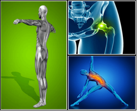
The hip joint is where the ball of the thigh bone (femur) joins the pelvis at a socket called the acetabulum. There is cartilage covering both the bone of the femur and the acetabulum of the pelvis in the hip joint. A joint lining tissue, called synovium, surrounds the hip joint. The synovium tissue produces fluid that lubricates the joint and provides nutrients to the cartilage of the joint. The ligaments around the hip joint attach the femur bone to the bony pelvis. There are a number of muscles and tendons that glide around the hip joint. Tiny fluid-filled sacs, called bursae, provide gliding surfaces for muscles and tendons around the hip joint. Major arteries and veins pass the front of the hip joint. The largest nerve of the body, the sciatic nerve, passes behind the hip joint.
If your hips are killing you, you probably spend a lot of time sitting – in the car, at work, on that spinning seat – which puts your hips in near-constant “flexion”, says Cori Lefkowith, NASM-certified personal trainer and owner of Redefining Strength in Orange County, California. Even running involves a repetitive flexion movement that can cause pain.

Approximately 15 degrees of hip extension is required to walk normally. If hip flexors are tight then in order to walk, compensatory movement needs to take place through the lower back causing back pain and premature disc degeneration. Like other joints, if we fail to take them through their full range on a regular basis we eventually lose mobility.
If you’re worried you’re headed toward a surgeon’s office, there might be hope. According to the Arthritis Foundation, the best way to avoid hip replacement surgery is to get active in an exercise program. In a study, people who participated in an exercise program for 12 weeks were 44 percent less likely to need joint-replacement surgery six years later than those who did not exercise.

Physician specialties that evaluate and treat low back pain range from generalists to subspecialists.These specialties include emergency medicine physicians, general medicine, family medicine, internal medicine, gynecology, spine surgeons (orthopaedics and neurosurgery), rheumatology, pain management, and physiatry. Other health care providers for low back pain include physical therapists, chiropractors, massage therapists, psychologists, and acupuncturists.

For those with pain localized to the lower back due to disc degeneration, fair evidence supports spinal fusion as equal to intensive physical therapy and slightly better than low-intensity nonsurgical measures.[15] Fusion may be considered for those with low back pain from acquired displaced vertebra that does not improve with conservative treatment,[14] although only a few of those who have spinal fusion experience good results.[15] There are a number of different surgical procedures to achieve fusion, with no clear evidence of one being better than the others.[83] Adding spinal implant devices during fusion increases the risks but provides no added improvement in pain or function.[11]
Moist heat may help relax your muscles. Put moist heat on the sore area for 10 to 15 minutes at a time before you do warm-up and stretching exercises. Moist heat includes heat patches or moist heating pads that you can buy at most drugstores, a wet washcloth or towel that has been heated in a microwave or the dryer, or a hot shower. Don’t use heat if you have swelling. 
The site navigation utilizes arrow, enter, escape, and space bar key commands. Left and right arrows move across top level links and expand / close menus in sub levels. Up and Down arrows will open main level menus and toggle through sub tier links. Enter and space open menus and escape closes them as well. Tab will move on to the next part of the site rather than go through menu items.
Consider working with a physical therapist to strengthen weak hip muscles. The physical therapists at Beacon Orthopaedists and Sports Medicine can also recommend a sport-specific training program. This is ideal for golfers, swimmers, runners, and any athlete who not only needs to preserve their hip but also strengthen it in order to remain competitive.
Squats. Using a squat machine will strengthen your quadriceps muscles on the front of your thigh and the hamstring muscles on the back of your thigh, both of which attach to your hip and give it support. The squat machine may be vertical, in which case you’ll start in a standing position and bend your knees until your thighs are parallel to the floor, or it may be on a sliding incline board.
Low back pain has been with humans since at least the Bronze Age. The oldest known surgical treatise – the Edwin Smith Papyrus, dating to about 1500 BCE – describes a diagnostic test and treatment for a vertebral sprain. Hippocrates (c. 460 BCE – c. 370 BCE) was the first to use a term for sciatic pain and low back pain; Galen (active mid to late second century CE) described the concept in some detail. Physicians through the end of the first millennium did not attempt back surgery and recommended watchful waiting. Through the Medieval period, folk medicine practitioners provided treatments for back pain based on the belief that it was caused by spirits.[99]
It is sometimes hard for an aggressive athlete to consider changing training schedules. It is also hard to accept the fact that a serious disease may exist. All athletes who suffer from low back pain should seek medical advice. Some situations might require reducing or stopping athletic activity until the problem is resolved. The body's ability to be active is worth preserving. 
Sacroiliac joint dysfunction. The sacroiliac joint connects the sacrum at the bottom of the spine to each side of the pelvis. It is a strong, low-motion joint that primarily absorbs shock and tension between the upper body and the lower body. The sacroiliac joint can become painful if it becomes inflamed (sacroiliitis) or if there is too much or too little motion of the joint.
Low back pain can be caused by tumors, either benign or malignant, that originate in the bone of the spine or pelvis and spinal cord (primary tumors) and those which originate elsewhere and spread to these areas (metastatic tumors). Symptoms range from localized pain to radiating severe pain and loss of nerve and muscle function (even incontinence of urine and stool) depending on whether or not the tumors affect the nervous tissue. Tumors of these areas are detected using imaging tests, such as plain X-rays, nuclear bone scanning, and CAT and MRI scanning.
Complaints of low back pain are one of the most common reasons people visit doctors.[9][42] For pain that has lasted only a few weeks, the pain is likely to subside on its own.[43] Thus, if a person's medical history and physical examination do not suggest a specific disease as the cause, medical societies advise against imaging tests such as X-rays, CT scans, and MRIs.[42] Individuals may want such tests but, unless red flags are present,[10][44] they are unnecessary health care.[9][43] Routine imaging increases costs, is associated with higher rates of surgery with no overall benefit,[45][46] and the radiation used may be harmful to one's health.[45] Fewer than 1% of imaging tests identify the cause of the problem.[9] Imaging may also detect harmless abnormalities, encouraging people to request further unnecessary testing or to worry.[9] Even so, MRI scans of the lumbar region increased by more than 300% among United States Medicare beneficiaries from 1994 to 2006.[11]
The hip is a basic ball-and-socket joint. The ball is the femoral head—a knob on the top of the thigh bone—and the socket is an indentation in the pelvic bone. There is cartilage lining the joint (called the labrum) and ligaments that attach the pelvic and thigh bones. Numerous muscles attach around the hip, too, moving the joint through the basic motions of flexion (bending), extension (extending the leg behind you), abduction (lifting the leg away from the body), adduction (moving the leg inward), internal rotation, and external rotation.
It is sometimes hard for an aggressive athlete to consider changing training schedules. It is also hard to accept the fact that a serious disease may exist. All athletes who suffer from low back pain should seek medical advice. Some situations might require reducing or stopping athletic activity until the problem is resolved. The body's ability to be active is worth preserving.
Workers who experience acute low back pain as a result of a work injury may be asked by their employers to have x-rays.[102] As in other cases, testing is not indicated unless red flags are present.[102] An employer's concern about legal liability is not a medical indication and should not be used to justify medical testing when it is not indicated.[102] There should be no legal reason for encouraging people to have tests which a health care provider determines are not indicated.[102]
Located deep in the front of the hip and connecting the leg, pelvis, and abdomen, the hip flexors— surprise, surprise— flex the hip. But despite being some of the most powerful muscles in our bodies (with a clearly important role), it’s easy to neglect our poor hip flexors— often without even knowing it. It turns out just working at a desk all day (guilty!) can really weaken hip flexors since they tend to shorten up while in a seated position. This tightness disrupts good posture and is a common cause of lower back pain. Weakened hip flexors can also increase the risk of foot, ankle, and knee injuries (especially among runners) Hip muscle weakness and overuse injuries in recreational runners. Niemuth, P.E., Johnson, R.J., Myers, M.J., et al. Rocky Mountain University of Health Professions, Provo, VT. Clinical Journal of Sport Medicine, 2005 Jan; 15 (1): 14-21.. So be sure to get up, stand up every hour or so! And giving the hip flexors some extra attention is not just about injury prevention. Adding power to workouts, working toward greater flexibility, and getting speedier while running is also, as they say, all in the hips The effect of walking speed on muscle function and mechanical energetics. Neptune, R.R., Sasaki, K., and Kautz, S.A. Department of Mechanical Engineering, The University of Texas, Austin, TX. Gait & Posture, 2008 Jul; 28 (1): 135-43.. 
Flexors are flexible muscle tissues that help a person stretch and move. The muscles that help you lift your knee toward your body and bend at the waist are known as the hip flexors. These muscles are located around the upper and inner things and pelvic region. Keeping the hip flexors strong is particularly important for active people and athletes.

So, who cares right? Wrong. Everyone has seen that little old man walking with a cane, hunched over almost to the point of staring at the ground. Do you think he always walked like that? I'd bet you he didn't. Maybe he had an injury that never healed properly, or maybe after spending years and years in a similar position, his body became tighter and tighter until eventually he ended up bent over.
So if you’re doing abs exercises and you feel like your hip flexors are putting in more work, refocus and dial in on the muscles you’re trying to target. It might sound trite to just “think” about a muscle working as you’re doing an exercise (for example, thinking about your abs contracting as you do a sit-up), but it might actually prevent you from mindlessly grinding out reps with poor form.
Following any period of prolonged inactivity, a regimen of low-impact exercises is advised. Speed walking, swimming, or stationary bike riding 30 minutes daily can increase muscle strength and flexibility. Yoga also can help stretch and strengthen muscles and improve posture. Consult a physician for a list of low-impact, age-appropriate exercises that are specifically targeted to strengthening lower back and abdominal muscles.
The AANS does not endorse any treatments, procedures, products or physicians referenced in these patient fact sheets. This information is provided as an educational service and is not intended to serve as medical advice. Anyone seeking specific neurosurgical advice or assistance should consult his or her neurosurgeon, or locate one in your area through the AANS’ Find a Board-certified Neurosurgeon” online tool.
Just because your hip flexor region feels sore doesn’t necessarily mean the muscles there are tight — in fact, they might need strengthening. This is where that sports science debate we mentioned earlier comes into play. It’s important to identify whether you’re tight or if the muscles are weak. Again, the Thomas Test will help you identify if you’re maybe stretching something that actually needs strengthening.
How to: Get into a high plank position on the floor, hands planted under your shoulders, butt down (a). Engage your abs by pulling your belly button in towards your spine (b). Squeeze your left glute to lift your left leg two inches off the ground, keeping your leg straight (c). Tap your left leg out to the side, then back to starting position. Repeat, then switch legs (d).
Behavioral therapy may be useful for chronic pain.[16] There are several types available, including operant conditioning, which uses reinforcement to reduce undesirable behaviors and increase desirable behaviors; cognitive behavioral therapy, which helps people identify and correct negative thinking and behavior; and respondent conditioning, which can modify an individual's physiological response to pain.[17] The benefit however is small.[91] Medical providers may develop an integrated program of behavioral therapies.[17] The evidence is inconclusive as to whether mindfulness-based stress reduction reduces chronic back pain intensity or associated disability, although it suggests that it may be useful in improving the acceptance of existing pain.[92][93]
Radicular pain. This type of pain can occur if a spinal nerve root becomes impinged or inflamed. Radicular pain may follow a nerve root pattern or dermatome down into the buttock and/or leg. Its specific sensation is sharp, electric, burning-type pain and can be associated with numbness or weakness (sciatica). It is typically felt on only one side of the body. 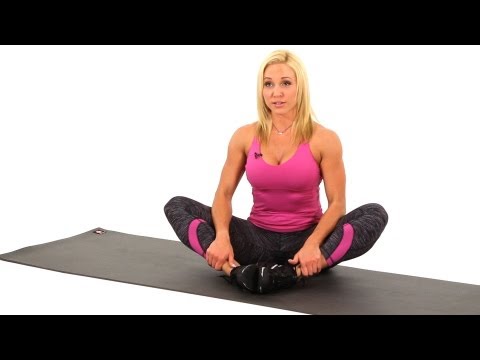
How to: Get into a high plank position on the floor, hands planted under your shoulders, butt down (a). Engage your abs by pulling your belly button in towards your spine (b). Squeeze your left glute to lift your left leg two inches off the ground, keeping your leg straight (c). Tap your left leg out to the side, then back to starting position. Repeat, then switch legs (d).
Degenerative disc disease. At birth, intervertebral discs are full of water and at their healthiest. As people age over time, discs lose hydration and wear down. As the disc loses hydration, it cannot resist forces as well, and transfers force to the disc wall that may develop tears and cause pain or weakening that can lead to a herniation. The disc can also collapse and contribute to stenosis.
References to any non-Onnit entity, product, service, person or source of information in this or any other Communication should not be considered an endorsement, either direct or implied, by the host, presenter or distributor of the Communication. The host(s), presenter(s) and/or distributor(s) of this Communication are not responsible for the content of any non-Onnit internet pages referenced in the Communication. Onnit is not liable or responsible for any advice, course of treatment, diagnosis or any other information or services you chose to follow without consulting a qualified medical professional. Before starting any new diet and/or exercise program, always be sure to check with your qualified medical professional.
Apply the above concept to your hips. When you sit, your hips are in a "flexed" position. Therefore, the muscles that flex your hips are in a shortened state. You probably spend at least a third of your day sitting down. Think about how much time those hip flexor muscles stay shortened. A lot. Over time, they become tighter and tighter until you look like the old man in the picture. So unless you want to look like that, perform the stretches shown below.
Putting the exercise in writing do not help me, I need to watch them doing them so, I can figer out how to do them, or if I should even try to do them. I use the flex extendors, lifting my legs one at a time from the flor to strengthen my thys, hip and buttox. And I try to remember to do the bridge excerise. I have had 2 total hip replacements , 7 months a part, in 2013. Trying to get stronger with cold weather will be 70 in Feb. Linda
Marvelously progressive, concise, and cogent guidelines for physicians on the treatment of low back pain. These guidelines almost entirely “get it right” in my opinion, and are completely consistent with recommendations I’ve been making for years on PainScience.com. They are particularly to be praised for strongly discouraging physicians from ordering imaging tests only “for patients with low back pain when severe or progressive neurologic deficits are present or when serious underlying conditions are suspected.”
There is a significant overlap of nerve supply to many of the discs, muscles, ligaments, and other spinal structures, and it can be difficult for the brain to accurately sense which is the cause of the pain. For example, a degenerated or torn lumbar disc can feel the same as a pulled muscle – both creating inflammation and painful muscle spasm in the same area. Muscles and ligaments heal rapidly, while a torn disc may or may not. The time course of pain helps determine the cause.
When was the last time you got on your gym's abductor or adductor machine and got in a good workout? It's probably been a while. Both are machines that don't get a lot of use, and they are often the target of coaches' ridicule on those "useless gym moves we should all skip" lists. Perhaps rightly so, especially if you're hopping on those machines hoping for a slimming effect.

Lumbar strain (acute, chronic): A lumbar strain is a stretch injury to the ligaments, tendons, and/or muscles of the low back. The stretching incident results in microscopic tears of varying degrees in these tissues. Lumbar strain is considered one of the most common causes of low back pain. The injury can occur because of overuse, improper use, or trauma. Soft-tissue injury is commonly classified as "acute" if it has been present for days to weeks. If the strain lasts longer than three months, it is referred to as "chronic." Lumbar strain most often occurs in people in their 40s, but it can happen at any age. The condition is characterized by localized discomfort in the low back area with onset after an event that mechanically stressed the lumbar tissues. The severity of the injury ranges from mild to severe, depending on the degree of strain and resulting spasm of the muscles of the low back. The diagnosis of lumbar strain is based on the history of injury, the location of the pain, and exclusion of nervous system injury. Usually, X-ray testing is only helpful to exclude bone abnormalities.
2016 — More editing, more! Added some better information about pain being a poor indicator, and the role of myofascial trigger points. This article has become extremely busy in the last couple months — about 4,000 readers per day, as described here — so I am really polishing it and making sure that it’s the best possible answer to people’s fears about back pain.

When you're working to strengthen the core, you'll want to focus on exercises that don't exacerbate lower back issues. "It's important to find out which movements (flexion, extension, rotation) cause pain or discomfort and to avoid those movements, while continuing to work into ranges that are not provoking," Dircksen says. Crockford suggests focusing on exercises that keep the core stable and avoiding twisting movements to avoid exacerbating pain.
The story of actor Andy Whitfield is a disturbing and educational example of a case that met these conditions — for sure the first two, and probably the third as well if we knew the details. Whitfield was the star of the hit TV show Spartacus (which is worthwhile, but rated very, very R17). The first sign of the cancer that killed him in 2011 was steadily worsening back pain. It’s always hard to diagnose a cancer that starts this way, but Whitfield was in the middle of intense physical training to look the part of history’s most famous gladiator. Back pain didn’t seem unusual at first, and some other symptoms may have been obscured. Weight loss could have even seemed like a training victory at first! It was many long months before he was diagnosed — not until the back pain was severe and constant. A scan finally revealed a large tumour pressing against his spine.

A sedentary lifestyle can lead to having weak and tight hip flexors as they are always in the shortened position. Tight hip flexors can lead to a limited range of motion, poor posture, lower back, and hip pain, and even injuries. These muscles need to get a workout when you are standing and doing movements such as raising your leg to climb stairs, run, or ride a bicycle.
Multiple sclerosis (MS) symptoms vary from person to person, and can last for days to months without periods of remission. Symptoms of MS include sexual problems and problems with the bowel, bladder, eyes, muscles, speech, swallowing, brain, and nervous system. The early symptoms and signs of multiple sclerosis usually start between age 20 and 40. MS in children, teens, and those over age 40 is rare. Treatment options for multiple sclerosis vary depending on the type and severity of symptoms. Medications may be prescribed to manage MS symptoms.
Strength training is another key part of the “do” category, Dr. Vasileff says. “It’s a good idea to focus on quad, hamstring, and glute strength,” he says. These muscles surround your hips and provide support, along with your core—which is another area to focus on. “Strengthening your core helps to normalize your walking pattern and stabilize how your pelvis and hips move,” Dr. Vasileff says. That translates to less pain and better hip mobility.

Really great content. I also had some lower back problem but now that I know the source, I will work on it. My counsins also talked to me about this product called Panifix, or "Unlock your hip flexor" which Gives You A Practical, Easy-to-follow Program You Can Use To Instantly Release Your Hip Flexors For More Strength, Better Health And All Day Energy. Proven Swipes And Creatives Here:https://tinyurl.com/yd6nbzfh

Hip hikers (also known as the pelvic drop) are great exercises to get your gluteal muscles working in a weight bearing position. To do the exercise, stand sideways with one foot on a step and the other hanging off. Keeping both knees straight, lower down your pelvis on one side so your foot moves toward the floor. Both knees should remain straight; the motion should come from your hip joint. Once your pelvis is lowered down, slowly raise it back up to the starting position. Repeat the exercise for 10 repetitions.
Iliopsoas syndrome, which is also called psoas syndrome or iliopsoas tendonitis, occurs when the iliopsoas muscles are injured. Lower back pain is the most common symptom; however, pain can also occur in the hip, thigh, or leg. The iliopsoas bursa, which is a fluid-filled sac located on the inside of the hip that reduces rubbing and friction, is also likely to become inflamed due to the proximity of the two structures. When this happens, the inflamed bursae will make it difficult to move.

Degenerative Conditions: Sometimes, degenerative conditions that are the normal result of aging may cause your low back pain. Conditions like spinal stenosis, arthritis, or degenerative disc disease can all cause pain. Congenital conditions, like spondylolisthesis or scoliosis, can also cause your back pain. For most degenerative back problems, movement and exercise have been proven to be effective in treating these conditions. A visit to your physical therapist can help you determine the correct progression of back exercises for your specific condition.

Just because your hip flexor region feels sore doesn’t necessarily mean the muscles there are tight — in fact, they might need strengthening. This is where that sports science debate we mentioned earlier comes into play. It’s important to identify whether you’re tight or if the muscles are weak. Again, the Thomas Test will help you identify if you’re maybe stretching something that actually needs strengthening.
The management of low back pain often includes medications for the duration that they are beneficial. With the first episode of low back pain the hope is a complete cure; however, if the problem becomes chronic, the goals may change to pain management and the recovery of as much function as possible. As pain medications are only somewhat effective, expectations regarding their benefit may differ from reality, and this can lead to decreased satisfaction.[13]
Other problems may occur along with low back pain. Chronic low back pain is associated with sleep problems, including a greater amount of time needed to fall asleep, disturbances during sleep, a shorter duration of sleep, and less satisfaction with sleep.[24] In addition, a majority of those with chronic low back pain show symptoms of depression[13] or anxiety.[17]
An injury to a ligament is called a sprain, and an injury to muscle or tendon is called a strain. Sprains and strains may be caused by repetitive movements or a single stressful incident. Symptoms and signs include pain and swelling. Though treatment depends upon the extent and location of the injury, rest, ice, compression, and elevation are key elements of treatment.
Avascular necrosis (also called osteonecrosis). This condition happens when blood flow to the hip bone slows and the bone tissue dies. Although it can affect other bones, avascular necrosis most often happens in the hip. It can be caused by a hip fracture or dislocation, or from the long-term use of high-dose steroids (such as prednisone), among other causes.
The bones of the hip are supported by specific muscles. The strongest muscle in the body is actually one of the main flexors of the hip, it is called the iliopsoas muscle. It has two attachments to the inner part of the hip as well as the lower part of the back. This muscle is involved in iliopsoas tendinitis, iliopsoas bursitis, and is one of the most common causes of muscle pain that causes hip pain. The tendons attach the muscles to the bones and the ligaments attach bone to bone. Areas that are often susceptible to increased pressure are the tendons or the muscles attached to the bone, so that is another source of pain in iliopsoas tendinitis. The bursa are pockets of fluid that also protect the top part of the bone to prevent pressure on the bone. There are about 160 bursa in the body, and when they get irritated, it can develop into bursitis.
Emerging technologies such as X-rays gave physicians new diagnostic tools, revealing the intervertebral disc as a source for back pain in some cases. In 1938, orthopedic surgeon Joseph S. Barr reported on cases of disc-related sciatica improved or cured with back surgery.[100] As a result of this work, in the 1940s, the vertebral disc model of low back pain took over,[99] dominating the literature through the 1980s, aiding further by the rise of new imaging technologies such as CT and MRI.[100] The discussion subsided as research showed disc problems to be a relatively uncommon cause of the pain. Since then, physicians have come to realize that it is unlikely that a specific cause for low back pain can be identified in many cases and question the need to find one at all as most of the time symptoms resolve within 6 to 12 weeks regardless of treatment.[99]
Imaging is indicated when there are red flags, ongoing neurological symptoms that do not resolve, or ongoing or worsening pain.[5] In particular, early use of imaging (either MRI or CT) is recommended for suspected cancer, infection, or cauda equina syndrome.[5] MRI is slightly better than CT for identifying disc disease; the two technologies are equally useful for diagnosing spinal stenosis.[5] Only a few physical diagnostic tests are helpful.[5] The straight leg raise test is almost always positive in those with disc herniation.[5] Lumbar provocative discography may be useful to identify a specific disc causing pain in those with chronic high levels of low back pain.[41] Similarly, therapeutic procedures such as nerve blocks can be used to determine a specific source of pain.[5] Some evidence supports the use of facet joint injections, transforminal epidural injections and sacroilliac injections as diagnostic tests.[5] Most other physical tests, such as evaluating for scoliosis, muscle weakness or wasting, and impaired reflexes, are of little use.[5]
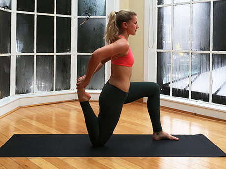
Start in a runner’s lunge with right leg forward, right knee over right ankle and back leg straight. Walk right foot over toward left hand, then drop right shin and thigh to the floor, making sure to keep right knee in line with right hip. Allow left leg to rest on the floor with top of left foot facing down. Take a moment to square your hips to the front of the room. Hold here, or hinge at hips and lower torso toward floor, allowing head to rest on forearms. Hold for at least 30 seconds, then repeat on opposite side. You want to feel a moderate stretch in the outside of the right thigh, but if this pose hurts your knees or feels too uncomfortable, stick with Thread the Needle.
In both younger and older patients, vertebral fractures take weeks to heal with rest and pain relievers. Compression fractures of vertebrae associated with osteoporosis can also be treated with a procedure called vertebroplasty or kyphoplasty, which can help to reduce pain. In this procedure, a balloon is inflated in the compressed vertebra, often returning some of its lost height. Subsequently, a "cement" (methymethacrylate) is injected into the balloon and remains to retain the structure and height of the body of the vertebra. Pain is relieved as the height of the collapsed vertebra is restored.
Low back pain that lasts at least one day and limits activity is a common complaint.[7] Globally, about 40% of people have LBP at some point in their lives,[7] with estimates as high as 80% of people in the developed world.[22] Approximately 9 to 12% of people (632 million) have LBP at any given point in time, and nearly one quarter (23.2%) report having it at some point over any one-month period.[7][8] Difficulty most often begins between 20 and 40 years of age.[1] Low back pain is more common among people aged 40–80 years, with the overall number of individuals affected expected to increase as the population ages.[7]
In the common presentation of acute low back pain, pain develops after movements that involve lifting, twisting, or forward-bending. The symptoms may start soon after the movements or upon waking up the following morning. The description of the symptoms may range from tenderness at a particular point to diffuse pain. It may or may not worsen with certain movements, such as raising a leg, or positions, such as sitting or standing. Pain radiating down the legs (known as sciatica) may be present. The first experience of acute low back pain is typically between the ages of 20 and 40. This is often a person's first reason to see a medical professional as an adult.[1] Recurrent episodes occur in more than half of people[23] with the repeated episodes being generally more painful than the first.[1]
Even though low back pain can sometimes be treated without major disruption to a person's life, athletes are often reluctant to seek medical help. Many of them deny or minimize complaints in order to avoid consequences, such as: having to decrease activity in order to recover, losing a position or being removed from a team, missing a competition, or letting the team down. Some athletes simply do not want to bother seeing a doctor for pain; they hope it will recover on its own.
Physical activity can also help you feel better. “Along with boosting your overall health, exercise can improve your OA symptoms” like pain, stiffness, fatigue, and even depression, says Leigh F. Callahan, PhD, associate director of the University of North Carolina Thurston Arthritis Research Center. One study found that people with knee OA who worked out regularly lowered their pain by 12% compared to those who didn’t.
It’s a common issue, says Prevention advisor Rob Danoff, director of family and emergency medicine residency programs at Aria Health in Philadelphia. "For people who sit a long time at work, the hip flexors and rotators become tight, and the gluteal muscles become weak," he says. "This combination negatively affects our ability to walk, maintain proper posture, and the stability of our spine."
Kneel on your mat with thighs perpendicular to the floor and tops of of your feet facing down. Place a yoga block between your feet. Bring your inner knees together. Slide your feet apart so they are slightly wider than your hips, and press the tops of your feet evenly into the mat. Slowly sit down on the yoga block. Use your hands to turn the top of your thighs inward. Allow the backs of your hands to rest on your thighs. Hold for at least 30 seconds.

These exercises can be done three to five times per week; be sure to build in a rest day here or there to allow your hip muscles to recover. Working to strengthen your knees and ankles can be done as well to be sure you completely work all muscles groups of your lower extremities. Remember, your ankle and knee muscles help control the position of your hips, just as your hip muscles control the position of your knees and ankles. They all work together in a kinetic chain.

Emerging technologies such as X-rays gave physicians new diagnostic tools, revealing the intervertebral disc as a source for back pain in some cases. In 1938, orthopedic surgeon Joseph S. Barr reported on cases of disc-related sciatica improved or cured with back surgery.[100] As a result of this work, in the 1940s, the vertebral disc model of low back pain took over,[99] dominating the literature through the 1980s, aiding further by the rise of new imaging technologies such as CT and MRI.[100] The discussion subsided as research showed disc problems to be a relatively uncommon cause of the pain. Since then, physicians have come to realize that it is unlikely that a specific cause for low back pain can be identified in many cases and question the need to find one at all as most of the time symptoms resolve within 6 to 12 weeks regardless of treatment.[99]

If you develop a sudden onset of low back pain, a visit to your physical therapist can help you determine the correct things to do to manage your acute pain. Your physical therapist should be able to analyze your lifestyle, movements, and overall medical history to help determine the likely cause of your pain. By focusing on these mechanical causes of back pain, you can make a change that may give you relief.
Like quadriceps, the hamstrings are 2-joint muscles. Unlike the quadriceps, though, the hamstrings reside at the back of your thigh. They attach at the siting bones, which are located on the underside of your pelvis. When the hamstring muscles contract, the effect is a pulling of the back of the pelvis down toward the back of the thigh, or a bringing of the lower extremity back behind you.
The AANS does not endorse any treatments, procedures, products or physicians referenced in these patient fact sheets. This information is provided as an educational service and is not intended to serve as medical advice. Anyone seeking specific neurosurgical advice or assistance should consult his or her neurosurgeon, or locate one in your area through the AANS’ Find a Board-certified Neurosurgeon” online tool.
THIS TOOL DOES NOT PROVIDE MEDICAL ADVICE. It is intended for general informational purposes only and does not address individual circumstances. It is not a substitute for professional medical advice, diagnosis or treatment and should not be relied on to make decisions about your health. Never ignore professional medical advice in seeking treatment because of something you have read on the WebMD Site. If you think you may have a medical emergency, immediately call your doctor or dial 911.
Avascular necrosis (also called osteonecrosis). This condition happens when blood flow to the hip bone slows and the bone tissue dies. Although it can affect other bones, avascular necrosis most often happens in the hip. It can be caused by a hip fracture or dislocation, or from the long-term use of high-dose steroids (such as prednisone), among other causes.

Management of low back pain depends on which of the three general categories is the cause: mechanical problems, non-mechanical problems, or referred pain.[52] For acute pain that is causing only mild to moderate problems, the goals are to restore normal function, return the individual to work, and minimize pain. The condition is normally not serious, resolves without much being done, and recovery is helped by attempting to return to normal activities as soon as possible within the limits of pain.[3] Providing individuals with coping skills through reassurance of these facts is useful in speeding recovery.[1] For those with sub-chronic or chronic low back pain, multidisciplinary treatment programs may help.[53] Initial management with non–medication based treatments is recommended, with NSAIDs used if these are not sufficiently effective.[6]
Infection of the discs (septic discitis) and bone (osteomyelitis) is extremely rare. These conditions lead to localized pain associated with fever. The bacteria found when these tissues are tested with laboratory cultures include Staphylococcus aureus and Mycobacterium tuberculosis (TB bacteria). TB infection in the spine is called Pott's disease. These are each very serious conditions requiring long courses of antibiotics. The sacroiliac joints rarely become infected with bacteria. Brucellosis is a bacterial infection that can involve the sacroiliac joints and is usually transmitted in raw goat's milk.
Nucleoplasty, also called plasma disc decompression (PDD), is a type of laser surgery that uses radiofrequency energy to treat people with low back pain associated with mildly herniated discs. Under x-ray guidance, a needle is inserted into the disc. A plasma laser device is then inserted into the needle and the tip is heated to 40-70 degrees Celsius, creating a field that vaporizes the tissue in the disc, reducing its size and relieving pressure on the nerves. Several channels may be made depending on how tissue needs to be removed to decompress the disc and nerve root.
Kneel on your mat with thighs perpendicular to the floor and tops of of your feet facing down. Place a yoga block between your feet. Bring your inner knees together. Slide your feet apart so they are slightly wider than your hips, and press the tops of your feet evenly into the mat. Slowly sit down on the yoga block. Use your hands to turn the top of your thighs inward. Allow the backs of your hands to rest on your thighs. Hold for at least 30 seconds.
A few cancers in their early stages can be hard to tell apart from ordinary back pain — a bone cancer in the vertebrae, for instance — and these create a frustrating diagnostic problem. They are too rare for doctors to inflict cancer testing on every low back pain patient “just in case.” And yet the possibility cannot be dismissed, either! It’s an unsolveable problem.
Following any period of prolonged inactivity, a regimen of low-impact exercises is advised. Speed walking, swimming, or stationary bike riding 30 minutes daily can increase muscle strength and flexibility. Yoga also can help stretch and strengthen muscles and improve posture. Consult a physician for a list of low-impact, age-appropriate exercises that are specifically targeted to strengthening lower back and abdominal muscles.
Knowing the exact source of your hip pain can provide you with peace of mind—but receiving the right treatment is even better. Let Dr. Hamilton provide you with both peace of mind and relief from your symptoms. Schedule an appointment with Dr. Hamilton today. For your convenience, he is available at Beacon East, Beacon West, or Summit Woods in Ohio as well as Beacon’s Northern Kentucky location.
Imaging is indicated when there are red flags, ongoing neurological symptoms that do not resolve, or ongoing or worsening pain.[5] In particular, early use of imaging (either MRI or CT) is recommended for suspected cancer, infection, or cauda equina syndrome.[5] MRI is slightly better than CT for identifying disc disease; the two technologies are equally useful for diagnosing spinal stenosis.[5] Only a few physical diagnostic tests are helpful.[5] The straight leg raise test is almost always positive in those with disc herniation.[5] Lumbar provocative discography may be useful to identify a specific disc causing pain in those with chronic high levels of low back pain.[41] Similarly, therapeutic procedures such as nerve blocks can be used to determine a specific source of pain.[5] Some evidence supports the use of facet joint injections, transforminal epidural injections and sacroilliac injections as diagnostic tests.[5] Most other physical tests, such as evaluating for scoliosis, muscle weakness or wasting, and impaired reflexes, are of little use.[5]
Stretching the hip muscles that sit on top of the bursae, part of the lining in your hip joint, can give you some relief from bursitis pain. Kneel on the leg that's giving you the pain, holding on to something sturdy for balance. Tilt your pelvis forward, tightening your gluteus muscles (the muscles in your buttocks). Then lean away from the side of your hip that hurts, for instance to the left if you're kneeling on your right knee. You should feel a stretch from the top of your hip bone down the side of your leg to your knee, Humphrey says. Hold the stretch for 30 seconds and repeat once or twice.
Magnetic resonance imaging (MRI) uses a magnetic force instead of radiation to create a computer-generated image. Unlike x-ray, which shows only bony structures, MRI scans also produce images of soft tissues such as muscles, ligaments, tendons, and blood vessels. An MRI may be ordered if a problem such as infection, tumor, inflammation, disc herniation or rupture, or pressure on a nerve is suspected. MRI is a noninvasive way to identify a condition requiring prompt surgical treatment. However, in most instances, unless there are “red flags” in the history or physical exam, an MRI scan is not necessary during the early phases of low back pain.