
Brace your core and bend your knees to lower down into a split squat. Your left knee should ideally form a 90-degree angle so that your thigh is parallel to the ground, and your right knee is hovering above the floor. (Quick position check: your left foot should be stepped out far enough that you can do this without letting your left knee go past your left toes—if you can't, hop your left foot out a bit farther away from the bench.)
The iliotibial band is a thickening of the fascia lata, the deep fascia of the thigh. Think of it as a thick long ligament like structure that connects the hip to the lower leg along the outside of the thigh. Tightness in the iliotibial band can cause patellofemoral pain, trochanteric bursitis, and friction syndromes at the knee. This is a hip stretch I commonly prescribe to runners and people suffering from knee pain.
Low back pain can be caused by tumors, either benign or malignant, that originate in the bone of the spine or pelvis and spinal cord (primary tumors) and those which originate elsewhere and spread to these areas (metastatic tumors). Symptoms range from localized pain to radiating severe pain and loss of nerve and muscle function (even incontinence of urine and stool) depending on whether or not the tumors affect the nervous tissue. Tumors of these areas are detected using imaging tests, such as plain X-rays, nuclear bone scanning, and CAT and MRI scanning.
How to do it: Stand with your feet together, holding dumbbells by your sides. Take a wide step out to your right and lower into a side lunge, reaching dumbbells on either side of right leg [as shown]. Bend your left knee and shift your weight into both legs, into a wide squat position, reaching the dumbbells to floor in front of you, then extend your right leg and shift your weight to the left, moving into a side lunge with your left leg.
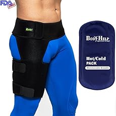
The JB Intensive Trainer Med Pro: A device that allows you to rehabilitate injury and pain, the JBIT MedPro helps strengthen hip muscles to alleviate pain over time. For older adults or those predisposed to joint and muscle conditions, the JBIT MedPro is an important preventative wellness solution, It can help mitigate the risk of worsened hip conditions that come with aging and wear and tear.
^ Enke, Oliver; New, Heather A.; New, Charles H.; Mathieson, Stephanie; McLachlan, Andrew J.; Latimer, Jane; Maher, Christopher G.; Lin, C.-W. Christine (2 July 2018). "Anticonvulsants in the treatment of low back pain and lumbar radicular pain: a systematic review and meta-analysis". Canadian Medical Association Journal. 190 (26): E786–E793. doi:10.1503/cmaj.171333. PMC 6028270. PMID 29970367.
4. Just swing it. For the front-to-back hip swing stretch, lie on the left side with hips stacked, propped up on the left elbow. Bend the left leg to a 90-degree angle and raise the right leg to hip level with toes pointed. Keep abs tight and swing the right leg all the way in front, then swing it all the way to the back, squeezing the booty along the way. Switch sides.
Your hip labrum is a band of cartilage-like tissue that courses around the outer rim of your hip socket. This labrum helps to support the joint and deepen the socket. Sometimes overuse or an injury to your hip can cause a tear in your labrum. A hip labrum tear may result in a condition called femoroacetabular impingement (FAI). When this happens, hip pain may result, and exercises to stretch and stabilize your hip may be performed.

Hamstring squeeze. Use the machine that works your hamstrings; you will either lie on your stomach or sit with a pad behind your knee. Push against the pad, moving your knee up toward the ceiling or backward (depending on which position you’re in). “In other words, bend your knees,” Pariser says. But to avoid cramps in your hamstring muscles, don’t bend your knee so much that your heels are too close to your buttocks. 
The National Institute of Neurological Disorders and Stroke (NINDS) is a component of the National Institutes of Health (NIH) and is the leading federal funder of research on disorders of the brain and nervous system. As a primary supporter of research on pain and pain mechanisms, NINDS is a member of the NIH Pain Consortium, which was established to promote collaboration among the many NIH Institutes and Centers with research programs and activities addressing pain. On an even broader scale, NIH participates in the Interagency Pain Research Coordinating Committee, a federal advisory committee that coordinates research across other U.S. Department of Health and Human Services agencies as well as the Departments of Defense and Veterans Affairs.
Of course, you know what it feels like to have a tight muscle. But tight hips aren't just uncomfortable—they can lead to all sorts of other aches and pains, especially in the lower back. "People focus on the hips and say their hips are tight, but we don't always think about the fact that the lower back connects to our legs at the hip," Charlee Atkins, C.S.C.S., instructor at Soul Annex in New York City and creator of Le Stretch class, tells SELF. Tight hip flexors make it harder for your pelvis to rotate properly, which can cause your lower back to overcompensate, "and this can be a setup for lower-back injury," Teo Mendez, M.D., an orthopedic surgeon at NY Orthopedics who focuses on operative and non-operative management of sports-related injuries, musculoskeletal injuries, and arthritis, tells SELF.
The outlook for low back pain absolutely depends on its precise cause. For example, acute strain injuries generally heal entirely with minimal treatment. On the other hand, bony abnormalities that are irritating the spinal cord can require significant surgical repair and the outlook depends on the surgical result. Long-term optimal results often involve exercise rehabilitation programs that can involve physical therapists.
Epidural injections of steroid drugs are frequently used to treat sciatica, despite limited evidence for their effectiveness. Moreover, these treatments are based on the assumption that reducing local inflammation in the vertebral column will relieve pain, but an association between structural abnormalities, inflammation, and sciatica symptoms has not been clearly demonstrated. NINDS-funded researchers are using a new imaging technique that can detect inflammation to better understand what causes chronic sciatica pain and to provide evidence to inform treatment selection.

Discography may be used when other diagnostic procedures fail to identify the cause of pain. This procedure involves the injection of a contrast dye into a spinal disc thought to be causing low back pain. The fluid’s pressure in the disc will reproduce the person’s symptoms if the disc is the cause. The dye helps to show the damaged areas on CT scans taken following the injection. Discography may provide useful information in cases where people are considering lumbar surgery or when their pain has not responded to conventional treatments.
Or anything else. Pain is a poor indicator, period! The human nervous system is really terrible about this: it routinely produces false alarms, and alarms that are much too loud. See Pain is Weird: Pain science reveals a volatile, misleading sensation that is often more than just a symptom, and sometimes worse than whatever started it. BACK TO TEXT
Lie on your back with your knees bent and feet flat on the floor. Place left ankle right below right knee, creating a “four” shape with left leg. Thread left arm through the opening you created with left leg and clasp hands behind right knee. Lift right foot off floor and pull right knee toward chest, flexing left foot. Hold for 30 seconds, then repeat on opposite side.
Hip pain is a common symptom experienced by many people. This condition can range from mild discomfort to severe pain that limits activities of daily living or functional mobility. Hip pain may prevent you from enjoying your normal work and recreational activities. When you visit a PT for hip pain, he or she will likely investigate the cause of your pain to help treat it properly.
Located deep in the front of the hip and connecting the leg, pelvis, and abdomen, the hip flexors— surprise, surprise— flex the hip. But despite being some of the most powerful muscles in our bodies (with a clearly important role), it’s easy to neglect our poor hip flexors— often without even knowing it. It turns out just working at a desk all day (guilty!) can really weaken hip flexors since they tend to shorten up while in a seated position. This tightness disrupts good posture and is a common cause of lower back pain. Weakened hip flexors can also increase the risk of foot, ankle, and knee injuries (especially among runners) Hip muscle weakness and overuse injuries in recreational runners. Niemuth, P.E., Johnson, R.J., Myers, M.J., et al. Rocky Mountain University of Health Professions, Provo, VT. Clinical Journal of Sport Medicine, 2005 Jan; 15 (1): 14-21.. So be sure to get up, stand up every hour or so! And giving the hip flexors some extra attention is not just about injury prevention. Adding power to workouts, working toward greater flexibility, and getting speedier while running is also, as they say, all in the hips The effect of walking speed on muscle function and mechanical energetics. Neptune, R.R., Sasaki, K., and Kautz, S.A. Department of Mechanical Engineering, The University of Texas, Austin, TX. Gait & Posture, 2008 Jul; 28 (1): 135-43..

Initial management with non–medication based treatments is recommended.[6] NSAIDs are recommended if these are not sufficiently effective.[6] Normal activity should be continued as much as the pain allows.[2] Medications are recommended for the duration that they are helpful.[13] A number of other options are available for those who do not improve with usual treatment. Opioids may be useful if simple pain medications are not enough, but they are not generally recommended due to side effects.[4][13] Surgery may be beneficial for those with disc-related chronic pain and disability or spinal stenosis.[14][15] No clear benefit has been found for other cases of non-specific low back pain.[14] Low back pain often affects mood, which may be improved by counseling or antidepressants.[13][16] Additionally, there are many alternative medicine therapies, including the Alexander technique and herbal remedies, but there is not enough evidence to recommend them confidently.[17] The evidence for chiropractic care[18] and spinal manipulation is mixed.[17][19][20][21]
The iliopsoas muscles are a group of two muscles—the psoas muscle and the iliacus muscle—located toward the front of the inner hip. The psoas muscles, in particular, is located in the lumbar (lower) region of the spine and extends through the pelvis to the femur. The iliopsoas muscles are the primary hip flexors, pulling the knee up off the ground when it contracts. Because the psoas muscle is also connected to the spine, it contributes to upright posture, assists in lumbar spine movement, and influences the spine’s curve.
Osteoarthritis. This condition results from wear and tear of the disc and facet joints. It causes pain, inflammation, instability, and stenosis to a variable degree, and can occur at a single level or multiple levels of the lower spine. Spinal osteoarthritis is associated with aging and is slowly progressive. It is also referred to as spondylosis or degenerative joint disease.
Congenital bone conditions: Congenital causes (existing from birth) of low back pain include scoliosis and spina bifida. Scoliosis is a sideways (lateral) curvature of the spine that can be caused when one lower extremity is shorter than the other (functional scoliosis) or because of an abnormal architecture of the spine (structural scoliosis). Children who are significantly affected by structural scoliosis may require treatment with bracing and/or surgery to the spine. Adults infrequently are treated surgically but often benefit by support bracing. Spina bifida is a birth defect in the bony vertebral arch over the spinal canal, often with absence of the spinous process. This birth defect most commonly affects the lowest lumbar vertebra and the top of the sacrum. Occasionally, there are abnormal tufts of hair on the skin of the involved area. Spina bifida can be a minor bony abnormality without symptoms. However, the condition can also be accompanied by serious nervous abnormalities of the lower extremities.

Before recommending exercises, physical therapists evaluate their patients to develop a routine that’s appropriate for their specific condition. Pariser says the following exercises, done at home and at the gym, are generally safe for everyone. “If a patient has already received a total hip replacement, however, certain precautions should be taken,” he says.
Consider working with a physical therapist to strengthen weak hip muscles. The physical therapists at Beacon Orthopaedists and Sports Medicine can also recommend a sport-specific training program. This is ideal for golfers, swimmers, runners, and any athlete who not only needs to preserve their hip but also strengthen it in order to remain competitive.
Sit on floor with knees bent and shins stacked with right leg on top. Use your hand to position right ankle on left knee. Ideally, the right knee will rest on the left thigh, but if your hips are tight, your right knee may point up toward the ceiling (overtime, as your hips become more open, your knee will lower). Keeping your hips squared to the front of the room, hinge at the hips and slowly walk hands slightly forward. If this is enough of a stretch, hold here, or fold your torso over your thighs to go deeper. Hold for at least 30 seconds, then repeat on opposite side.

Coccydynia is an inflammation of the bony area (tailbone or coccyx) located between the buttocks. Coccydynia is associated with pain and tenderness at the tip of the tailbone between the buttocks. Pain is often worsened by sitting. There are many causes of tailbone pain that can mimic coccydynia including: fracture, pilonidal cysts, infection, and sciatica. Treatment methods include medication and rest.
Intradiscal electrothermal therapy (IDET) is a treatment for discs that are cracked or bulging as a result of degenerative disc disease. The procedure involves inserting a catheter through a small incision at the site of the disc in the back. A special wire is passed through the catheter and an electrical current is applied to heat the disc, which helps strengthen the collagen fibers of the disc wall, reducing the bulging and the related irritation of the spinal nerve. IDET is of questionable benefit.
This Web site provides general educational information on health-related issues and provides access to health-related resources for the convenience of our users. This site and its health-related information and resources are not a substitute for professional medical advice or for the care that patients receive from their physicians or other health care providers.
The use of lumbar supports in the form of wide elastic bands that can be tightened to provide support to the lower back and abdominal muscles to prevent low back pain remains controversial. Such supports are widely used despite a lack of evidence showing that they actually prevent pain. Multiple studies have determined that the use of lumbar supports provides no benefit in terms of the prevention and treatment of back pain. Although there have been anecdotal case reports of injury reduction among workers using lumbar support belts, many companies that have back belt programs also have training and ergonomic awareness programs. The reported injury reduction may be related to a combination of these or other factors. Furthermore, some caution is advised given that wearing supportive belts may actually lead to or aggravate back pain by causing back muscles to weaken from lack of use.
The treatment of lumbar strain consists of resting the back (to avoid reinjury), medications to relieve pain and muscle spasm, local heat applications, massage, and eventual (after the acute episode resolves) reconditioning exercises to strengthen the low back and abdominal muscles. Initial treatment at home might include heat application, acetaminophen (Tylenol) or ibuprofen (Advil, Motrin), and avoiding reinjury and heavy lifting. Prescription medications that are sometimes used for acute low back pain include anti-inflammatory medications, such as sulindac (Clinoril), naproxen (Naprosyn), and ketorolac (Toradol) by injection or by mouth, muscle relaxants, such as carisoprodol (Soma), cyclobenzaprine (Flexeril), methocarbamol (Robaxin), and metaxalone (Skelaxin), as well as analgesics, such as tramadol (Ultram).

In the vast majority of patients with low back pain, symptoms can be attributed to nonspecific mechanical factors. However, in a much smaller percentage of patients, the cause of back pain may be something more serious, such as cancer, cauda equina syndrome, spinal infection, spinal compression fractures, spinal stress fractures, ankylosing spondylitis, or aneurysm.
^ Machado, GC; Maher, CG; Ferreira, PH; Pinheiro, MB; Lin, CW; Day, RO; McLachlan, AJ; Ferreira, ML (31 March 2015). "Efficacy and safety of paracetamol for spinal pain and osteoarthritis: systematic review and meta-analysis of randomised placebo controlled trials". BMJ (Clinical Research Ed.). 350: h1225. doi:10.1136/bmj.h1225. PMC 4381278. PMID 25828856.
Sit on floor with knees bent and shins stacked with right leg on top. Use your hand to position right ankle on left knee. Ideally, the right knee will rest on the left thigh, but if your hips are tight, your right knee may point up toward the ceiling (overtime, as your hips become more open, your knee will lower). Keeping your hips squared to the front of the room, hinge at the hips and slowly walk hands slightly forward. If this is enough of a stretch, hold here, or fold your torso over your thighs to go deeper. Hold for at least 30 seconds, then repeat on opposite side.
There is a significant overlap of nerve supply to many of the discs, muscles, ligaments, and other spinal structures, and it can be difficult for the brain to accurately sense which is the cause of the pain. For example, a degenerated or torn lumbar disc can feel the same as a pulled muscle – both creating inflammation and painful muscle spasm in the same area. Muscles and ligaments heal rapidly, while a torn disc may or may not. The time course of pain helps determine the cause.

Magnetic resonance imaging (MRI) uses a magnetic force instead of radiation to create a computer-generated image. Unlike x-ray, which shows only bony structures, MRI scans also produce images of soft tissues such as muscles, ligaments, tendons, and blood vessels. An MRI may be ordered if a problem such as infection, tumor, inflammation, disc herniation or rupture, or pressure on a nerve is suspected. MRI is a noninvasive way to identify a condition requiring prompt surgical treatment. However, in most instances, unless there are “red flags” in the history or physical exam, an MRI scan is not necessary during the early phases of low back pain.
Gait analysis studies in the elderly show that they typically have a shortened step length. Whether that is a result of tight hip flexors or due to reduced balance, the propensity to walk with shorter steps will itself lead to tightness in hip flexors and anterior joint structures. Hip stretches may be a relatively easy preventative strategy for the elderly with gait abnormalities and may help to prevent falls.
Iliopsoas syndrome, which is also called psoas syndrome or iliopsoas tendonitis, occurs when the iliopsoas muscles are injured. Lower back pain is the most common symptom; however, pain can also occur in the hip, thigh, or leg. The iliopsoas bursa, which is a fluid-filled sac located on the inside of the hip that reduces rubbing and friction, is also likely to become inflamed due to the proximity of the two structures. When this happens, the inflamed bursae will make it difficult to move.

