
The lumbar spine (lower back) consists of five vertebrae in the lower part of the spine between the ribs and the pelvis. The bones (vertebrae) that form the spine in the back are cushioned by small discs. These discs are round and flat, with a tough, outer layer (annulus) that surrounds a jellylike material called the nucleus. Located between each of the vertebra in the spinal column, discs act as shock absorbers for the spinal bones. Thick ligaments attached to the vertebrae hold the pulpy disc material in place. Of the 31 pairs of spinal nerves and roots, five lumbar (L1-L5) and five sacral (S1-S5) nerve pairs connect beginning in the area of the lower back.
Low back pain results in large economic costs. In the United States, it is the most common type of pain in adults, responsible for a large number of missed work days, and is the most common musculoskeletal complaint seen in the emergency department.[25] In 1998, it was estimated to be responsible for $90 billion in annual health care costs, with 5% of individuals incurring most (75%) of the costs.[25] Between 1990 and 2001 there was a more than twofold increase in spinal fusion surgeries in the US, despite the fact that there were no changes to the indications for surgery or new evidence of greater usefulness.[11] Further costs occur in the form of lost income and productivity, with low back pain responsible for 40% of all missed work days in the United States.[101] Low back pain causes disability in a larger percentage of the workforce in Canada, Great Britain, the Netherlands and Sweden than in the US or Germany.[101]
Just because your hip flexor region feels sore doesn’t necessarily mean the muscles there are tight — in fact, they might need strengthening. This is where that sports science debate we mentioned earlier comes into play. It’s important to identify whether you’re tight or if the muscles are weak. Again, the Thomas Test will help you identify if you’re maybe stretching something that actually needs strengthening.
Most functional exercises—ones that mimic everyday movements such as squats, hip hinges (deadlifts, for example), lunges, steps-ups—stretch and strengthen your hip muscles in some way. So if you strength train and do a variety of these sorts of movements, you're probably working these important muscles without even realizing it. On the other hand, if you mostly focus on exercise methods that have you doing the same movement over and over again, like running or cycling, there's a good chance your hips aren't as strong as they should be. And that can have a negative impact on not only your workouts, but how you move through life in general.
But moving is important for hip and knee OA. It causes your joints to compress and release, bringing blood flow, nutrients, and oxygen into the cartilage. “This can help prolong the function and longevity of your joints,” says Eric Robertson, DPT, a physical therapist and associate professor of clinical physical therapy at the University of Southern California.
Pain along the inside of the hip may be due to tendinitis or strain of the adductor muscles. Adductors (or inner thigh muscles) pull the leg inward as it is moving forward—the faster the movement, the greater the degree of adduction. Since footprints of a runner are almost single file as opposed to the side-by-side footprints of a walker, there is some degree of adduction occurring during running.
The management of low back pain often includes medications for the duration that they are beneficial. With the first episode of low back pain the hope is a complete cure; however, if the problem becomes chronic, the goals may change to pain management and the recovery of as much function as possible. As pain medications are only somewhat effective, expectations regarding their benefit may differ from reality, and this can lead to decreased satisfaction.[13]
Electrodiagnostics are procedures that, in the setting of low back pain, are primarily used to confirm whether a person has lumbar radiculopathy. The procedures include electromyography (EMG), nerve conduction studies (NCS), and evoked potential (EP) studies. EMG assesses the electrical activity in a muscle and can detect if muscle weakness results from a problem with the nerves that control the muscles. Very fine needles are inserted in muscles to measure electrical activity transmitted from the brain or spinal cord to a particular area of the body. NCSs are often performed along with EMG to exclude conditions that can mimic radiculopathy. In NCSs, two sets of electrodes are placed on the skin over the muscles. The first set provides a mild shock to stimulate the nerve that runs to a particular muscle. The second set records the nerve’s electrical signals, and from this information nerve damage that slows conduction of the nerve signal can be detected. EP tests also involve two sets of electrodes—one set to stimulate a sensory nerve, and the other placed on the scalp to record the speed of nerve signal transmissions to the brain.

Bony encroachment: Any condition that results in movement or growth of the vertebrae of the lumbar spine can limit the space (encroachment) for the adjacent spinal cord and nerves. Causes of bony encroachment of the spinal nerves include foraminal narrowing (narrowing of the portal through which the spinal nerve passes from the spinal column, out of the spinal canal to the body, commonly as a result of arthritis), spondylolisthesis (slippage of one vertebra relative to another), and spinal stenosis (compression of the nerve roots or spinal cord by bony spurs or other soft tissues in the spinal canal). Spinal-nerve compression in these conditions can lead to sciatica pain that radiates down the lower extremities. Spinal stenosis can cause lower-extremity pains that worsen with walking and are relieved by resting (mimicking the pains of poor circulation). Treatment of these afflictions varies, depending on their severity, and ranges from rest and exercises to epidural cortisone injections and surgical decompression by removing the bone that is compressing the nervous tissue.
The lower back where most back pain occurs includes the five vertebrae (referred to as L1-L5) in the lumbar region, which supports much of the weight of the upper body. The spaces between the vertebrae are maintained by round, rubbery pads called intervertebral discs that act like shock absorbers throughout the spinal column to cushion the bones as the body moves. Bands of tissue known as ligaments hold the vertebrae in place, and tendons attach the muscles to the spinal column. Thirty-one pairs of nerves are rooted to the spinal cord and they control body movements and transmit signals from the body to the brain.
Quick anatomy lesson. When we talk about the hips, we're talking about any muscle that crosses over the hip joint, says Laura Miranda D.P.T., M.S.P.T., C.S.C.S., a New York City-based trainer and creator of the Pursuit training program. Which, there are many, including all of the glute muscles, the hamstrings, the inner thigh muscles, and the psoas muscles (deep core muscles that attach your pelvis to your spine). Each of these muscles has some specific roles, but overall, the hip muscles stabilize your pelvis and thighbone as you move. They also allow you to bend at the hips, lift your legs out to the side (abduct), and bring your legs back in toward one another (adduct). Basically, they do a lot, and when they're weak or tight or otherwise not working in an optimal way, you can not only end up with cranky hips, but other body parts may overcompensate and take on too much work—leaving you with other, seemingly unrelated, issues, like knee pain.
^ Paige, Neil M.; Miake-Lye, Isomi M.; Booth, Marika Suttorp; Beroes, Jessica M.; Mardian, Aram S.; Dougherty, Paul; Branson, Richard; Tang, Baron; Morton, Sally C.; Shekelle, Paul G. (11 April 2017). "Association of Spinal Manipulative Therapy With Clinical Benefit and Harm for Acute Low Back Pain". JAMA. 317 (14): 1451–1460. doi:10.1001/jama.2017.3086. PMC 5470352. PMID 28399251.
Of course, you know what it feels like to have a tight muscle. But tight hips aren't just uncomfortable—they can lead to all sorts of other aches and pains, especially in the lower back. "People focus on the hips and say their hips are tight, but we don't always think about the fact that the lower back connects to our legs at the hip," Charlee Atkins, C.S.C.S., instructor at Soul Annex in New York City and creator of Le Stretch class, tells SELF. Tight hip flexors make it harder for your pelvis to rotate properly, which can cause your lower back to overcompensate, "and this can be a setup for lower-back injury," Teo Mendez, M.D., an orthopedic surgeon at NY Orthopedics who focuses on operative and non-operative management of sports-related injuries, musculoskeletal injuries, and arthritis, tells SELF.
For strains and tendinitis at the top of the hamstrings, treatment is the same as that used for hip flexor problems. Hamstring stretching and strengthening—such as side lunges, inward leg raises, and backward leg raises—is important. Deep tissue massage may also be beneficial, but in general, this is a difficult problem that usually takes a while to resolve.
Imaging is indicated when there are red flags, ongoing neurological symptoms that do not resolve, or ongoing or worsening pain.[5] In particular, early use of imaging (either MRI or CT) is recommended for suspected cancer, infection, or cauda equina syndrome.[5] MRI is slightly better than CT for identifying disc disease; the two technologies are equally useful for diagnosing spinal stenosis.[5] Only a few physical diagnostic tests are helpful.[5] The straight leg raise test is almost always positive in those with disc herniation.[5] Lumbar provocative discography may be useful to identify a specific disc causing pain in those with chronic high levels of low back pain.[41] Similarly, therapeutic procedures such as nerve blocks can be used to determine a specific source of pain.[5] Some evidence supports the use of facet joint injections, transforminal epidural injections and sacroilliac injections as diagnostic tests.[5] Most other physical tests, such as evaluating for scoliosis, muscle weakness or wasting, and impaired reflexes, are of little use.[5]
Low back pain may be classified based on the signs and symptoms. Diffuse pain that does not change in response to particular movements, and is localized to the lower back without radiating beyond the buttocks, is classified as nonspecific, the most common classification.[5] Pain that radiates down the leg below the knee, is located on one side (in the case of disc herniation), or is on both sides (in spinal stenosis), and changes in severity in response to certain positions or maneuvers is radicular, making up 7% of cases.[5] Pain that is accompanied by red flags such as trauma, fever, a history of cancer or significant muscle weakness may indicate a more serious underlying problem and is classified as needing urgent or specialized attention.[5]
I think you should mention that for some people, stretching is not the solution and that it will deteriorate their posture. Some people need stretching, but most people I know need to strengthen their "overstretched" hip flexors. Many people can't do a single hanging leg raise. Check this site if you want to know more about the importance of hip flexors bit.ly/Unlock_Your_Hip_Flexor Report

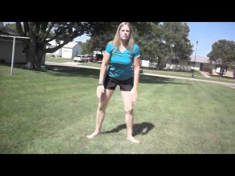
Hip flexors. These hardworking muscles are crucial in foundational movements such as sitting, standing, walking and running — they act as a bridge connecting your torso to your lower body. Some muscles in this group can be notoriously weak or tight and those of you who have ever had issues with this part of your body will know the uncomfortable pain of either all too well. There’s a lot of debate in the world of sports science over how much you should strengthen and stretch your hip flexors — we’ll explain.
In this study, one patient with sciatica was sent for ten MRIs, which produced 49 distinct “findings,” 16 of them unique, none of which occurred in all ten reports. On average, each radiologist made about a dozen errors, seeing one or two things that weren’t there and missing about ten things that were. Yikes. Read a more detailed and informal description of this study.
When you tell your doctor your hip hurts, the first thing she should do is confirm that your hip is actually the problem. Women might say they have hip pain, but what they may mean is that they have pain in the side of the upper thigh or upper buttock, or they may be experiencing lower back pain, says Stephanie E. Siegrist, MD, an orthopedic surgeon in Rochester, New York, and a spokeswoman for the American Academy of Orthopaedic Surgeons. Hip pain is often felt in the groin or on the outside of the hip directly over where the hip joint (a ball-and-socket joint) is located.
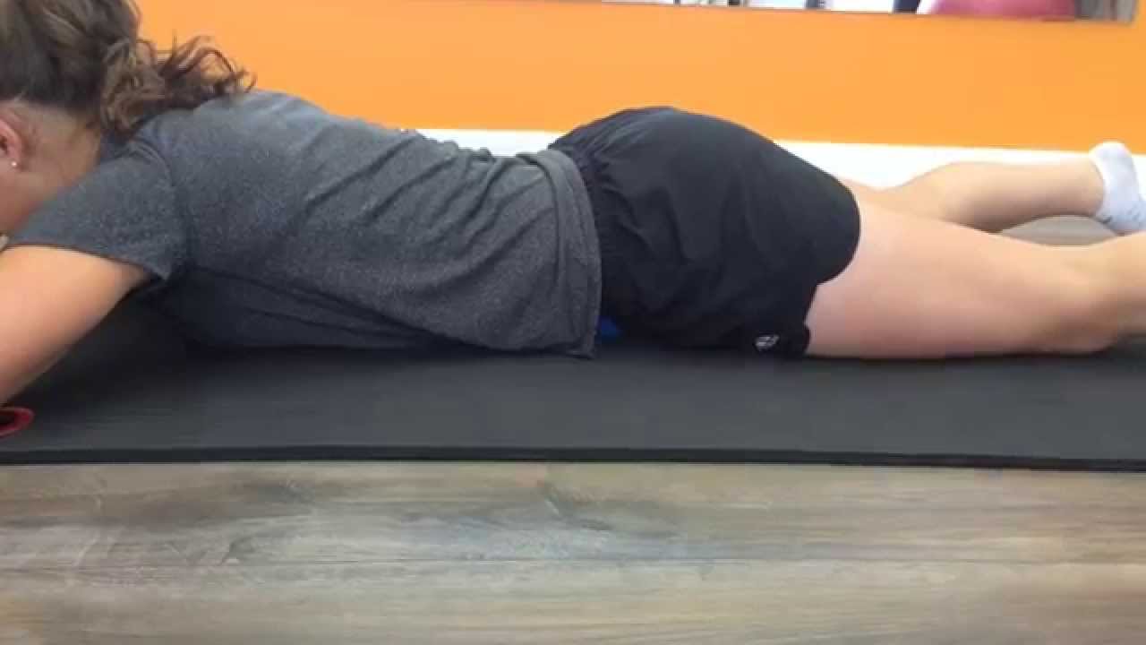
Bridge: Still lying on your back with your feet flat on floor, lift your hips and torso off the floor into a bridge. Then interlace your hands underneath your hips and press your shoulders and upper arms into the floor, lifting your hips higher. Hold for 10 seconds. Lower yourself slowly back down, rolling down from the top of your spine to your tailbone. Repeat three times.
Spondylolisthesis. This condition occurs when one vertebra slips over the adjacent one. There are 5 types of spondylolisthesis but the most common are secondary to a defect or fracture of the pars (between the facet joints) or mechanical instability of the facet joints (degenerative). The pain can be caused by instability (back) or compression of the nerves (leg).

Nerve irritation: The nerves of the lumbar spine can be irritated by mechanical pressure (impingement) by bone or other tissues, or from disease, anywhere along their paths -- from their roots at the spinal cord to the skin surface. These conditions include lumbar disc disease (radiculopathy), bony encroachment, and inflammation of the nerves caused by a viral infection (shingles). See descriptions of these conditions below.
The site navigation utilizes arrow, enter, escape, and space bar key commands. Left and right arrows move across top level links and expand / close menus in sub levels. Up and Down arrows will open main level menus and toggle through sub tier links. Enter and space open menus and escape closes them as well. Tab will move on to the next part of the site rather than go through menu items.

Kidneys — The kidneys are a matched pair. One painful kidney can cause back pain on one side or the other. Kidney pain can feel like back pain, and may occur on only one side. It is usually quite lateral, and just barely low enough to qualify as “low” back pain. However, when kidney stones descend through the ureters, they can cause (terrible) pain in the low back. Kidney stone pain is often so severe and develops so rapidly that it isn’t mistaken for a back pain problem.

Endometriosis (when the uterus lining grows somewhere else) can cause pelvic tenderness, which some women describe as hip pain. Pain from the back and spine also can be felt around the buttocks and hip, Siegrist says. Sciatica, a pinched nerve, typically affects one side of the body and can cause pain in the back of the right or left hip — the pain from sciatica can start in your lower back and travel down to your buttocks and legs.
Low back pain (LBP) is a common disorder involving the muscles, nerves, and bones of the back.[4] Pain can vary from a dull constant ache to a sudden sharp feeling.[4] Low back pain may be classified by duration as acute (pain lasting less than 6 weeks), sub-chronic (6 to 12 weeks), or chronic (more than 12 weeks).[3] The condition may be further classified by the underlying cause as either mechanical, non-mechanical, or referred pain.[5] The symptoms of low back pain usually improve within a few weeks from the time they start, with 40–90% of people completely better by six weeks.[2]
