
Bone scans are used to detect and monitor infection, fracture, or disorders in the bone. A small amount of radioactive material is injected into the bloodstream and will collect in the bones, particularly in areas with some abnormality. Scanner-generated images can be used to identify specific areas of irregular bone metabolism or abnormal blood flow, as well as to measure levels of joint disease.
A pinched nerve causes pain, numbness, or tingling in the affected area due to pressure on a nerve. Caral tunnel and sciatica are two examples of conditions caused by a pinched nerve. A pinched nerve is diagnosed by taking a patient history and performing a physical examination. Electromyography may be performed. Treatment for a pinched nerve depends on the underlying cause.
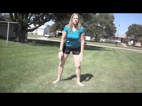
Lay on your back on your mat and pull your knees to your chest. Place your hands on the inside arches of your feet and open your knees wider than shoulder-width apart. Keeping your back pressed into the mat as much as possible, press your feet into hands while pulling down on feet, creating resistance. Breathe deeply and hold for at least 30 seconds.

Medicine FinderLatest NewsSlip, slap, slop early to save your skinComplementary medicine users have worse cancer survivalVideo: BoredomPut down the tablet; pick up a bookVideo: Lift your mood with some weightsQuicken your step to live longerThis web site is intended for Australian residents and is not a substitute for independent professional advice. Information and interactions contained in this Web site are for information purposes only and are not intended to be used to diagnose, treat, cure or prevent any disease. Further, the accuracy, currency and completeness of the information available on this Web site cannot be guaranteed. Tonic Digital Media Pty Ltd, its affiliates and their respective servants and agents do not accept any liability for any injury, loss or damage incurred by use of or reliance on the information made available via or through myDr whether arising from negligence or otherwise. See Privacy Policy and Disclaimer.2001-2019 myDr.com.au © | All Rights Reserved About UsContact UsDisclaimerPrivacy PolicyAdvertising PolicySitemap

The hip joint is where the ball of the thigh bone (femur) joins the pelvis at a socket called the acetabulum. There is cartilage covering both the bone of the femur and the acetabulum of the pelvis in the hip joint. A joint lining tissue, called synovium, surrounds the hip joint. The synovium tissue produces fluid that lubricates the joint and provides nutrients to the cartilage of the joint. The ligaments around the hip joint attach the femur bone to the bony pelvis. There are a number of muscles and tendons that glide around the hip joint. Tiny fluid-filled sacs, called bursae, provide gliding surfaces for muscles and tendons around the hip joint. Major arteries and veins pass the front of the hip joint. The largest nerve of the body, the sciatic nerve, passes behind the hip joint.
Veritas Health publishes original and accessible health related content written by more than 100 physician authors and peer-reviewed by a 16 member Medical Advisory Board. The Veritas Health platform comprising of Spine-health.com, Arthritis-health.com, Sports-health.com, and Pain-health.com, provides comprehensive information on back pain, arthritis, sports injuries, and chronic pain conditions. For more information visit Veritashealth.com.

Degenerative Conditions: Sometimes, degenerative conditions that are the normal result of aging may cause your low back pain. Conditions like spinal stenosis, arthritis, or degenerative disc disease can all cause pain. Congenital conditions, like spondylolisthesis or scoliosis, can also cause your back pain. For most degenerative back problems, movement and exercise have been proven to be effective in treating these conditions. A visit to your physical therapist can help you determine the correct progression of back exercises for your specific condition.
Low back pain may be classified based on the signs and symptoms. Diffuse pain that does not change in response to particular movements, and is localized to the lower back without radiating beyond the buttocks, is classified as nonspecific, the most common classification.[5] Pain that radiates down the leg below the knee, is located on one side (in the case of disc herniation), or is on both sides (in spinal stenosis), and changes in severity in response to certain positions or maneuvers is radicular, making up 7% of cases.[5] Pain that is accompanied by red flags such as trauma, fever, a history of cancer or significant muscle weakness may indicate a more serious underlying problem and is classified as needing urgent or specialized attention.[5]
Back “spasms” are a largely a myth — there’s no such thing a sustained painful contractions of muscles in otherwise healthy people (see Cramps, Spasms, Tremors & Twitches) — but the kernel of truth in the idea of “spasms” may be the idea of trigger points, which are hypothetical “micro cramps,” tiny patches of painfully contracting muscle. Although this idea is controversial, it is nevertheless one of the most likely explanations for common aches and pains that mostly stick to one area (especially the back) and have no other obvious cause. See Back Pain & Trigger Points. BACK TO TEXT

Non-mechanical Disease Processes: Sometimes, non-mechanical disease processes like cancer, kidney stones, or a tumor may cause low back pain. These symptoms are usually, but not always, accompanied by other symptoms like unexpected weight loss, fever, or malaise that indicate a non-mechanical cause of your pain. These diseases are rare, but they can happen, so if your back pain continues for more than a few weeks after physical therapy treatment begins, a visit to your doctor is certainly in order to rule out a sinister problem.
It is sometimes hard for an aggressive athlete to consider changing training schedules. It is also hard to accept the fact that a serious disease may exist. All athletes who suffer from low back pain should seek medical advice. Some situations might require reducing or stopping athletic activity until the problem is resolved. The body's ability to be active is worth preserving.

Chronic back pain is defined as pain that persists for 12 weeks or longer, even after an initial injury or underlying cause of acute low back pain has been treated. About 20 percent of people affected by acute low back pain develop chronic low back pain with persistent symptoms at one year. In some cases, treatment successfully relieves chronic low back pain, but in other cases pain persists despite medical and surgical treatment.
Doing the bridge exercise in the morning gets your muscles working, activated, and engaged and will help support you the rest of the day, says Humphrey. Lie on your back with your legs bent and your feet flat on the floor, hip-width apart. Press down through your ankles and raise your buttocks off the floor while you tighten your abdominal muscles. Keep your knees aligned with your ankles and aim for a straight line from knees to shoulders, being sure not to arch your back; hold this position for three to five seconds and then slowly lower your buttocks back to the floor. Start with one set of 10 and build up to two or three sets.

Vertebroplasty and kyphoplasty are minimally invasive treatments to repair compression fractures of the vertebrae caused by osteoporosis. Vertebroplasty uses three-dimensional imaging to assist in guiding a fine needle through the skin into the vertebral body, the largest part of the vertebrae. A glue-like bone cement is then injected into the vertebral body space, which quickly hardens to stabilize and strengthen the bone and provide pain relief. In kyphoplasty, prior to injecting the bone cement, a special balloon is inserted and gently inflated to restore height to the vertebral structure and reduce spinal deformity.

