
Vertebroplasty and kyphoplasty are minimally invasive treatments to repair compression fractures of the vertebrae caused by osteoporosis. Vertebroplasty uses three-dimensional imaging to assist in guiding a fine needle through the skin into the vertebral body, the largest part of the vertebrae. A glue-like bone cement is then injected into the vertebral body space, which quickly hardens to stabilize and strengthen the bone and provide pain relief. In kyphoplasty, prior to injecting the bone cement, a special balloon is inserted and gently inflated to restore height to the vertebral structure and reduce spinal deformity.

You’d think so. But consider this story of a motorcycle accident: many years ago, a friend hit a car that had pulled out from a side street. He flew over the car & landed on his head. Bystanders showed their ignorance of spinal fracture by, yikes, carelessly moving him. In fact, his thoracic spine was significantly fractured … yet the hospital actually refused to do an X-ray because he had no obvious symptoms of a spinal fracture. Incredible! The next day, a horrified orthopedic surgeon ordered an X-ray immediately, confirming the fracture & quite possibly saved him from paralysis.
Long periods of inactivity in bed are no longer recommended, as this treatment may actually slow recovery. Spinal manipulation for periods of up to one month has been found to be helpful in some patients who do not have signs of nerve irritation. Future injury is avoided by using back-protection techniques during activities and support devices as needed at home or work.

^ Jump up to: a b c d American College of Occupational and Environmental Medicine (February 2014), "Five Things Physicians and Patients Should Question", Choosing Wisely: an initiative of the ABIM Foundation, American College of Occupational and Environmental Medicine, archived from the original on 11 September 2014, retrieved 24 February 2014, which cites
Model Heather Lin grew up in the deep south but is currently hustling in New York, working at a bank. Whether she is biking home from work, deadlifting, kicking a heavy bag, or pouring all of her effort into a bootcamp class, it's important to her to find time in her busy day to work out. She feels her best when she is strong and energized, and blogs about her health and fitness journey at The Herbivore Warrior.
Pain is generally an unpleasant feeling in response to an event that either damages or can potentially damage the body's tissues. There are four main steps in the process of feeling pain: transduction, transmission, perception, and modulation.[12] The nerve cells that detect pain have cell bodies located in the dorsal root ganglia and fibers that transmit these signals to the spinal cord.[33] The process of pain sensation starts when the pain-causing event triggers the endings of appropriate sensory nerve cells. This type of cell converts the event into an electrical signal by transduction. Several different types of nerve fibers carry out the transmission of the electrical signal from the transducing cell to the posterior horn of spinal cord, from there to the brain stem, and then from the brain stem to the various parts of the brain such as the thalamus and the limbic system. In the brain, the pain signals are processed and given context in the process of pain perception. Through modulation, the brain can modify the sending of further nerve impulses by decreasing or increasing the release of neurotransmitters.[12]
^ Jump up to: a b c d e f Qaseem, A; Wilt, TJ; McLean, RM; Forciea, MA; Clinical Guidelines Committee of the American College of, Physicians. (4 April 2017). "Noninvasive Treatments for Acute, Subacute, and Chronic Low Back Pain: A Clinical Practice Guideline From the American College of Physicians". Annals of Internal Medicine. 166 (7): 514–530. doi:10.7326/M16-2367. PMID 28192789.
Traction involves the use of weights and pulleys to apply constant or intermittent force to gradually “pull” the skeletal structure into better alignment. Some people experience pain relief while in traction, but that relief is usually temporary. Once traction is released the back pain tends to return. There is no evidence that traction provides any longterm benefits for people with low back pain.

Walking is an easy way to strengthen your bones and muscles, burn calories, and lift your mood. If you don't currently walk for exercise, try walking for five to 10 minutes every day. Gradually build up to 30 minutes a day. It's easy to sneak walking into your day. Take up golf, window-shop at a large mall, or visit a walking-only destination, such as a museum or botanical garden.
Stand tall with your hips square and bend your right knee, bringing your foot towards your bum. Grab the right foot with your right hand and actively pull the foot closer to your glutes. As you do this, send the right knee down towards the ground and keep both knees together. squeeze your butt to promote a posterior pelvic tilt and hold — then switch sides.
There are many additional sources of pain, including claudication pain (from stenosis) myelopathic pain, neuropathic pain, deformity, tumors, infections, pain from inflammatory conditions (such as rheumatoid arthritis or ankylosing spondylitis), and pain that originates from another part of the body and presents in the lower back (such as kidney stones, or ulcerative colitis).
There are a number of ways to classify low back pain with no consensus that any one method is best.[5] There are three general types of low back pain by cause: mechanical back pain (including nonspecific musculoskeletal strains, herniated discs, compressed nerve roots, degenerative discs or joint disease, and broken vertebra), non-mechanical back pain (tumors, inflammatory conditions such as spondyloarthritis, and infections), and referred pain from internal organs (gallbladder disease, kidney stones, kidney infections, and aortic aneurysm, among others).[5] Mechanical or musculoskeletal problems underlie most cases (around 90% or more),[5][34] and of those, most (around 75%) do not have a specific cause identified, but are thought to be due to muscle strain or injury to ligaments.[5][34] Rarely, complaints of low back pain result from systemic or psychological problems, such as fibromyalgia and somatoform disorders.[34]
Our Keep-It™ guarantee is valid for the first-time purchase of a formula, and redeemable up to three months (90 days) after the purchase date. Multiple bottles, foods, apparel and gear do not fall under this guarantee, however, they may be applicable for return. Fitness equipment, personal care products, knowledge purchases, and DVDs are not eligible for return or refund. For more information and a full list of products that qualify, visit our Keep-It™ page. Further details can be found on our Refund Policy support page.
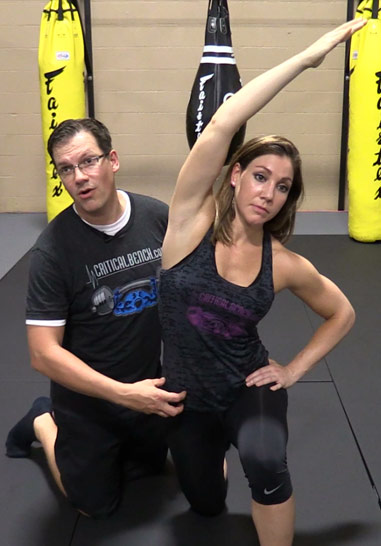
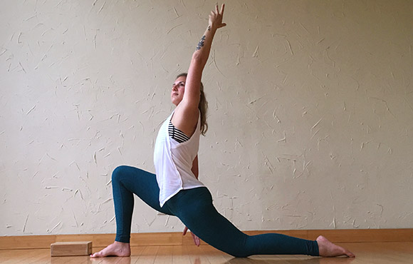
Hip flexors. These hardworking muscles are crucial in foundational movements such as sitting, standing, walking and running — they act as a bridge connecting your torso to your lower body. Some muscles in this group can be notoriously weak or tight and those of you who have ever had issues with this part of your body will know the uncomfortable pain of either all too well. There’s a lot of debate in the world of sports science over how much you should strengthen and stretch your hip flexors — we’ll explain.
Apply the above concept to your hips. When you sit, your hips are in a "flexed" position. Therefore, the muscles that flex your hips are in a shortened state. You probably spend at least a third of your day sitting down. Think about how much time those hip flexor muscles stay shortened. A lot. Over time, they become tighter and tighter until you look like the old man in the picture. So unless you want to look like that, perform the stretches shown below.
When we stand, the lower back is functioning to support the weight of the upper body. When we bend, extend, or rotate at the waist, the lower back is involved in the movement. Therefore, injury to the structures important for weight bearing, such as the bony spine, muscles, tendons, and ligaments, often can be detected when the body is standing erect or used in various movements.
In the common presentation of acute low back pain, pain develops after movements that involve lifting, twisting, or forward-bending. The symptoms may start soon after the movements or upon waking up the following morning. The description of the symptoms may range from tenderness at a particular point to diffuse pain. It may or may not worsen with certain movements, such as raising a leg, or positions, such as sitting or standing. Pain radiating down the legs (known as sciatica) may be present. The first experience of acute low back pain is typically between the ages of 20 and 40. This is often a person's first reason to see a medical professional as an adult.[1] Recurrent episodes occur in more than half of people[23] with the repeated episodes being generally more painful than the first.[1]
Foraminotomy is an operation that “cleans out” or enlarges the bony hole (foramen) where a nerve root exits the spinal canal. Bulging discs or joints thickened with age can cause narrowing of the space through which the spinal nerve exits and can press on the nerve, resulting in pain, numbness, and weakness in an arm or leg. Small pieces of bone over the nerve are removed through a small slit, allowing the surgeon to cut away the blockage and relieve pressure on the nerve.
Sacroiliac joint dysfunction. The sacroiliac joint connects the sacrum at the bottom of the spine to each side of the pelvis. It is a strong, low-motion joint that primarily absorbs shock and tension between the upper body and the lower body. The sacroiliac joint can become painful if it becomes inflamed (sacroiliitis) or if there is too much or too little motion of the joint.
Radiofrequency denervation is a procedure using electrical impulses to interrupt nerve conduction (including the conduction of pain signals). Using x-ray guidance, a needle is inserted into a target area of nerves and a local anesthetic is introduced as a way of confirming the involvement of the nerves in the person’s back pain. Next, the region is heated, resulting in localized destruction of the target nerves. Pain relief associated with the technique is temporary and the evidence supporting this technique is limited.
Flexors are flexible muscle tissues that help a person stretch and move. The muscles that help you lift your knee toward your body and bend at the waist are known as the hip flexors. These muscles are located around the upper and inner things and pelvic region. Keeping the hip flexors strong is particularly important for active people and athletes.
Like quadriceps, the hamstrings are 2-joint muscles. Unlike the quadriceps, though, the hamstrings reside at the back of your thigh. They attach at the siting bones, which are located on the underside of your pelvis. When the hamstring muscles contract, the effect is a pulling of the back of the pelvis down toward the back of the thigh, or a bringing of the lower extremity back behind you.
You’d think so. But consider this story of a motorcycle accident: many years ago, a friend hit a car that had pulled out from a side street. He flew over the car & landed on his head. Bystanders showed their ignorance of spinal fracture by, yikes, carelessly moving him. In fact, his thoracic spine was significantly fractured … yet the hospital actually refused to do an X-ray because he had no obvious symptoms of a spinal fracture. Incredible! The next day, a horrified orthopedic surgeon ordered an X-ray immediately, confirming the fracture & quite possibly saved him from paralysis.
Avascular necrosis (also called osteonecrosis). This condition happens when blood flow to the hip bone slows and the bone tissue dies. Although it can affect other bones, avascular necrosis most often happens in the hip. It can be caused by a hip fracture or dislocation, or from the long-term use of high-dose steroids (such as prednisone), among other causes.
Paget's disease can be diagnosed on plain X-rays. However, a bone biopsy is occasionally necessary to ensure the accuracy of the diagnosis. Bone scanning is helpful to determine the extent of the disease, which can involve more than one bone area. A blood test, alkaline phosphatase, is useful for diagnosis andmonitoring response to therapy. Treatment options include aspirin, other anti-inflammatory medicines, pain medications, and medications that slow therate of bone turnover, such as calcitonin (Calcimar, Miacalcin), etidronate (Didronel), alendronate (Fosamax), risedronate (Actonel), and pamidronate (Aredia).
Your hip labrum is a band of cartilage-like tissue that courses around the outer rim of your hip socket. This labrum helps to support the joint and deepen the socket. Sometimes overuse or an injury to your hip can cause a tear in your labrum. A hip labrum tear may result in a condition called femoroacetabular impingement (FAI). When this happens, hip pain may result, and exercises to stretch and stabilize your hip may be performed.
Fitness level: Back pain is more common among people who are not physically fit. Weak back and abdominal muscles may not properly support the spine. “Weekend warriors”—people who go out and exercise a lot after being inactive all week—are more likely to suffer painful back injuries than people who make moderate physical activity a daily habit. Studies show that low-impact aerobic exercise is beneficial for the maintaining the integrity of intervertebral discs.
Grade II (moderate): A larger tear in your muscle that makes it difficult to move and causes a moderate amount of pain, especially when you move the affected muscle, swelling, and tenderness. You may have 5 percent to 50 percent loss of function and you may be limping. You can't go back to sporting activities until the tear is completely healed. These injuries can take anywhere from a couple weeks to a few months to heal, depending on how bad they are.


Avoiding injury to the low back is a method of preventing low back pain. Additionally, conditioning exercise programs designed to strengthen the lumbar area and adjacent tissues can help to minimize risk of injury to the low back. Specific programs to relieve and prevent back pain can be designed with the help of physical therapists and other treating health-care professionals.
Really great content. I also had some lower back problem but now that I know the source, I will work on it. My counsins also talked to me about this product called Panifix, or "Unlock your hip flexor" which Gives You A Practical, Easy-to-follow Program You Can Use To Instantly Release Your Hip Flexors For More Strength, Better Health And All Day Energy. Proven Swipes And Creatives Here:https://tinyurl.com/yd6nbzfh

Pain is generally an unpleasant feeling in response to an event that either damages or can potentially damage the body's tissues. There are four main steps in the process of feeling pain: transduction, transmission, perception, and modulation.[12] The nerve cells that detect pain have cell bodies located in the dorsal root ganglia and fibers that transmit these signals to the spinal cord.[33] The process of pain sensation starts when the pain-causing event triggers the endings of appropriate sensory nerve cells. This type of cell converts the event into an electrical signal by transduction. Several different types of nerve fibers carry out the transmission of the electrical signal from the transducing cell to the posterior horn of spinal cord, from there to the brain stem, and then from the brain stem to the various parts of the brain such as the thalamus and the limbic system. In the brain, the pain signals are processed and given context in the process of pain perception. Through modulation, the brain can modify the sending of further nerve impulses by decreasing or increasing the release of neurotransmitters.[12]
Avoid stress: Sometimes the worst thing about pain is the stress it can cause. If you aren’t aching all over you can spend most of your time worrying the pain will return or, if you are already in pain, you can start to stress over how it is impacting your life. You really should try to avoid working yourself into a panic though – it really isn’t doing you any good.
Age: The first attack of low back pain typically occurs between the ages of 30 and 50, and back pain becomes more common with advancing age. As people grow older, loss of bone strength from osteoporosis can lead to fractures, and at the same time, muscle elasticity and tone decrease. The intervertebral discs begin to lose fluid and flexibility with age, which decreases their ability to cushion the vertebrae. The risk of spinal stenosis also increases with age.
Those are some great stretches! I own a personal training studio in Severna Park, Maryland. Majority of my clients have physical limitations – so it’s important for them to stay flexible. I send these to my clients and even do these exercises for myself. I highly recommend these stretches to anyone, even people without physical limitations. I love the fact these are actually videos and not just stretches because it’s so much easier for people to figure out how to perform the stretches. You guys are the real MVP!
Discectomy (the partial removal of a disc that is causing leg pain) can provide pain relief sooner than nonsurgical treatments.[14] Discectomy has better outcomes at one year but not at four to ten years.[14] The less invasive microdiscectomy has not been shown to result in a different outcome than regular discectomy.[14] For most other conditions, there is not enough evidence to provide recommendations for surgical options.[14] The long-term effect surgery has on degenerative disc disease is not clear.[14] Less invasive surgical options have improved recovery times, but evidence regarding effectiveness is insufficient.[14]
In terms of diagnosing hip pain, typically a patient will expect when they come in to be asked about their symptoms, and it’s very important to find out when did these symptoms start, how long they have been going on, how frequent they are, if they come on in the morning or the evening, do they come on with any certain activity, and if there is something that makes it better or worse. The intensity of the pain is also important. Does it have any associated radiating symptoms? Is it localized in one spot or does it move? After getting a history and finding out what type of pain the patient is having, which also includes whether the pain is dull, aching, sharp, or intense, then it’s important to do a good physical exam. The physical examination involves testing the muscle strength, testing for sensation, doing provocative maneuvers which might help us rule out one type of injury from another.


