
Following any period of prolonged inactivity, a regimen of low-impact exercises is advised. Speed walking, swimming, or stationary bike riding 30 minutes daily can increase muscle strength and flexibility. Yoga also can help stretch and strengthen muscles and improve posture. Consult a physician for a list of low-impact, age-appropriate exercises that are specifically targeted to strengthening lower back and abdominal muscles. 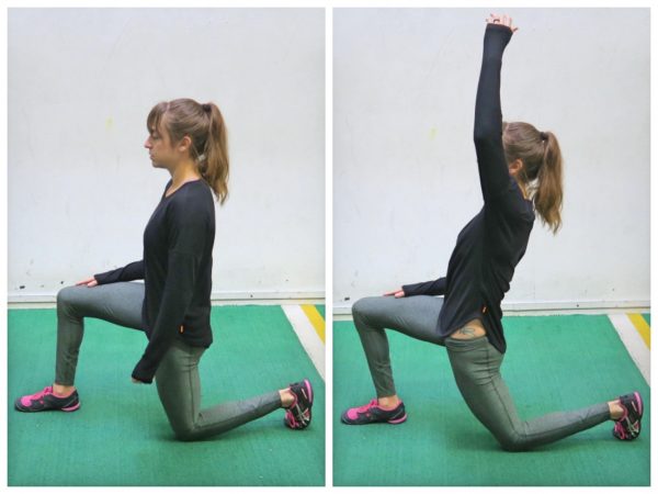
Icing a joint that’s inflamed because of arthritis or bursitis can lower inflammation and help with hip pain. “If it’s very painful, I sometimes tell patients to ice four or five times daily for about 10 to 15 minutes,” says Amy Humphrey, DPT, a physical therapist at Body Dynamics, Inc. in Arlington, Va. Use an ice pack, wrap a towel around it, and put it where you feel the pain.

Since the hip flexors are so easy to overuse and strain, it is important to stretch them before exercise or strenuous activity. Because of their connectedness to other muscle groups in the legs (like the quadriceps), it is important to stretch the muscle group as a whole, not individually. This ensures that the entire area is warmed up and ready for the strain of exercise or activity, not just one or two individual muscles.
Analgesic medications are those specifically designed to relieve pain. They include OTC acetaminophen and aspirin, as well as prescription opioids such as codeine, oxycodone, hydrocodone, and morphine. Opioids should be used only for a short period of time and under a physician’s supervision. People can develop a tolerance to opioids and require increasingly higher dosages to achieve the same effect. Opioids can also be addictive. Their side effects can include drowsiness, constipation, decreased reaction time, and impaired judgment. Some specialists are concerned that chronic use of opioids is detrimental to people with back pain because they can aggravate depression, leading to a worsening of the pain.
While leg lifts, certain ab exercises, and even hula hooping can all help work the hips, the hip flexors can still be a tricky part of the body to stretch Kinetics of hula hooping: An inverse dynamics analysis. Cluff, T., Robertson, D.G., and Balasubramaniam, R. School of Human Kinetics, University of Ottawa, Ottawa, Ontario, Canada. Human Movement Science, 2008 Aug; 27 (4): 622-35.. To get them even stronger and more flexible, try these five simple hip flexor stretches:


Premkumar et al present evidence that the traditional “red flags” for ominous causes of back pain can be quite misleading. The correlation between red flags and ominous diagnoses is poor, and prone to producing false negatives: that is, no red flags even when there is something more serious than unexplained pain going on. In a survey of almost 10,000 patients “the absence of red flag responses did not meaningfully decrease the likelihood of a red flag diagnosis.“ This is not even remotely a surprise to anyone who paid attention in back pain school, but it’s good to have some harder data on it.
"Lower back pain is the most common musculoskeletal ailment in the U.S., and can often be mitigated by strengthening the core musculature," Blake Dircksen, D.P.T., C.S.C.S., a physical therapist at Bespoke Treatments New York, tells SELF. "The 'core' is a cylinder of abdominal and back muscles that wraps around the body like a corset," Dircksen explains. (The glutes are also considered a part of the core, since they connect to the pelvis and ultimately the back and abdominal muscles.) As with any muscles, by strengthening them, you will increase the amount of weight your lower back can comfortably move, which means it will be better equipped to handle the same stress from your workouts and everyday life without getting as achey.
How to: Stand tall, feet slightly wider than shoulder-width apart, toes pointed out at 45-degree angles (a). Keep your back straight, knees over toes and your weight in the heels of your feet (b). Engage your glutes and thighs as you lower into a deep squat until thighs parallel to the ground (or as close as you can get them) (c). Powering through your heels, push up to return to starting position (d). Repeat.
Low back pain can be caused by tumors, either benign or malignant, that originate in the bone of the spine or pelvis and spinal cord (primary tumors) and those which originate elsewhere and spread to these areas (metastatic tumors). Symptoms range from localized pain to radiating severe pain and loss of nerve and muscle function (even incontinence of urine and stool) depending on whether or not the tumors affect the nervous tissue. Tumors of these areas are detected using imaging tests, such as plain X-rays, nuclear bone scanning, and CAT and MRI scanning.

Discography may be used when other diagnostic procedures fail to identify the cause of pain. This procedure involves the injection of a contrast dye into a spinal disc thought to be causing low back pain. The fluid’s pressure in the disc will reproduce the person’s symptoms if the disc is the cause. The dye helps to show the damaged areas on CT scans taken following the injection. Discography may provide useful information in cases where people are considering lumbar surgery or when their pain has not responded to conventional treatments.
I am a science writer, former massage therapist, and I was the assistant editor at ScienceBasedMedicine.org for several years. I have had my share of injuries and pain challenges as a runner and ultimate player. My wife and I live in downtown Vancouver, Canada. See my full bio and qualifications, or my blog, Writerly. You might run into me on Facebook or Twitter.

Most functional exercises—ones that mimic everyday movements such as squats, hip hinges (deadlifts, for example), lunges, steps-ups—stretch and strengthen your hip muscles in some way. So if you strength train and do a variety of these sorts of movements, you're probably working these important muscles without even realizing it. On the other hand, if you mostly focus on exercise methods that have you doing the same movement over and over again, like running or cycling, there's a good chance your hips aren't as strong as they should be. And that can have a negative impact on not only your workouts, but how you move through life in general.
Stand tall with your hips square and bend your right knee, bringing your foot towards your bum. Grab the right foot with your right hand and actively pull the foot closer to your glutes. As you do this, send the right knee down towards the ground and keep both knees together. squeeze your butt to promote a posterior pelvic tilt and hold — then switch sides.
Pain along the inside of the hip may be due to tendinitis or strain of the adductor muscles. Adductors (or inner thigh muscles) pull the leg inward as it is moving forward—the faster the movement, the greater the degree of adduction. Since footprints of a runner are almost single file as opposed to the side-by-side footprints of a walker, there is some degree of adduction occurring during running.
In both younger and older patients, vertebral fractures take weeks to heal with rest and pain relievers. Compression fractures of vertebrae associated with osteoporosis can also be treated with a procedure called vertebroplasty or kyphoplasty, which can help to reduce pain. In this procedure, a balloon is inflated in the compressed vertebra, often returning some of its lost height. Subsequently, a "cement" (methymethacrylate) is injected into the balloon and remains to retain the structure and height of the body of the vertebra. Pain is relieved as the height of the collapsed vertebra is restored.

The bony lumbar spine is designed so that vertebrae "stacked" together can provide a movable support structure while also protecting the spinal cord from injury. The spinal cord is composed of nervous tissue that extends down the spinal column from the brain. Each vertebra has a spinous process, a bony prominence behind the spinal cord, which shields the cord's nervous tissue from impact trauma. Vertebrae also have a strong bony "body" (vertebral body) in front of the spinal cord to provide a platform suitable for weight bearing of all tissues above the buttocks. The lumbar vertebrae stack immediately atop the sacrum bone that is situated in between the buttocks. On each side, the sacrum meets the iliac bone of the pelvis to form the sacroiliac joints of the buttocks.
Start kneeling on your mat with knees hip-width apart and hips directly over knees. Press your shins and the tops of your feet into the mat. Bring your hands to your low back, fingers pointing down, and rest palms above glutes. Inhale and lift your chest, and then slowly start to lean your torso back. From here, bring your right hand to rest on your right heel and then your left hand to your left heel. (If you can't reach your heels, turn your toes under; it will be easier to reach your heels in this modification.) Press your thighs forward so they are perpendicular to the floor. Keep your head in a relatively neutral position or, if it doesn't strain your neck, drop it back. Hold for 30 seconds. To come out of the pose, bring your hands to your hips and slowly, leading with your chest, lift your torso as you press the thighs down toward the floor.
Swimming is a low impact form of exercise that can help to relieve the pressure on your joints. It’s thought that water aerobics may help to reduce the impact on your joints by up to 75%!2 Be careful though; start with a beginner’s class and always do only what you’re comfortable with – try to avoid over-exercising or pushing your muscles too far!
Spinal manipulation and spinal mobilization are approaches in which professionally licensed specialists (doctors of chiropractic care) use their hands to mobilize, adjust, massage, or stimulate the spine and the surrounding tissues. Manipulation involves a rapid movement over which the individual has no control; mobilization involves slower adjustment movements. The techniques have been shown to provide small to moderate short-term benefits in people with chronic low back pain. Evidence supporting their use for acute or subacute low back pain is generally of low quality. Neither technique is appropriate when a person has an underlying medical cause for the back pain such as osteoporosis, spinal cord compression, or arthritis.
Compressive pain is a result of pressure or irritation on the spinal cord, nerves that leave the spine. For example, if an intervertebral disc herniates (usually called a ruptured disc) and pushes into the spinal canal, it can cause problems with the nerve. Usually this pressure or irritation causes pain, numbness, and muscle weakness where the nerve travels.
However, even the things you do every day — like sitting in front of a computer or at a desk for hours — can both weaken and shorten (tighten) your hip flexors, making them more prone to injury. Because of this, exercises (such as squats) and targeted stretches which focus on strengthening the hip muscles and improving hip mobility are key to preventing injuries.
Overall, the outcome for acute low back pain is positive. Pain and disability usually improve a great deal in the first six weeks, with complete recovery reported by 40 to 90%.[2] In those who still have symptoms after six weeks, improvement is generally slower with only small gains up to one year. At one year, pain and disability levels are low to minimal in most people. Distress, previous low back pain, and job satisfaction are predictors of long-term outcome after an episode of acute pain.[2] Certain psychological problems such as depression, or unhappiness due to loss of employment may prolong the episode of low back pain.[13] Following a first episode of back pain, recurrences occur in more than half of people.[23]
Simply stand up straight with your feet about shoulder-width apart. Slowly bend your knees and hips, lowering yourself until your knees obscure your toes or you achieve a 90 degree angle. Hold for a count of 5 and then gently resume your original position. This can be a tough one so again, don’t overdo it and hold on to a table if you need a little extra support! Try to repeat between 5-10 times.

