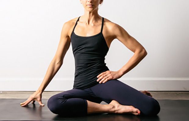
Model Heather Lin grew up in the deep south but is currently hustling in New York, working at a bank. Whether she is biking home from work, deadlifting, kicking a heavy bag, or pouring all of her effort into a bootcamp class, it's important to her to find time in her busy day to work out. She feels her best when she is strong and energized, and blogs about her health and fitness journey at The Herbivore Warrior.
2016 — More editing, more! Added some better information about pain being a poor indicator, and the role of myofascial trigger points. This article has become extremely busy in the last couple months — about 4,000 readers per day, as described here — so I am really polishing it and making sure that it’s the best possible answer to people’s fears about back pain.
Veritas Health publishes original and accessible health related content written by more than 100 physician authors and peer-reviewed by a 16 member Medical Advisory Board. The Veritas Health platform comprising of Spine-health.com, Arthritis-health.com, Sports-health.com, and Pain-health.com, provides comprehensive information on back pain, arthritis, sports injuries, and chronic pain conditions. For more information visit Veritashealth.com.

But moving is important for hip and knee OA. It causes your joints to compress and release, bringing blood flow, nutrients, and oxygen into the cartilage. “This can help prolong the function and longevity of your joints,” says Eric Robertson, DPT, a physical therapist and associate professor of clinical physical therapy at the University of Southern California.
MRI and x-ray for low back pain are surprisingly unreliable,1 because things like bulging discs usually aren’t a deal,2 most back pain goes away on its own,3 and trigger points (“muscle knots”) are common and can be alarmingly intense but aren’t dangerous.4 Most patients are much better off when they feel confident about these things. The power of justified, rational confidence is a huge factor in back pain.5 Sadly, many healthcare professionals continue to perpetuate the idea of fragile backs,6 which undermines that valuable confidence.

Start kneeling on your mat with knees hip-width apart and hips directly over knees. Press your shins and the tops of your feet into the mat. Bring your hands to your low back, fingers pointing down, and rest palms above glutes. Inhale and lift your chest, and then slowly start to lean your torso back. From here, bring your right hand to rest on your right heel and then your left hand to your left heel. (If you can't reach your heels, turn your toes under; it will be easier to reach your heels in this modification.) Press your thighs forward so they are perpendicular to the floor. Keep your head in a relatively neutral position or, if it doesn't strain your neck, drop it back. Hold for 30 seconds. To come out of the pose, bring your hands to your hips and slowly, leading with your chest, lift your torso as you press the thighs down toward the floor.

Or anything else. Pain is a poor indicator, period! The human nervous system is really terrible about this: it routinely produces false alarms, and alarms that are much too loud. See Pain is Weird: Pain science reveals a volatile, misleading sensation that is often more than just a symptom, and sometimes worse than whatever started it. BACK TO TEXT
Low back pain may be classified based on the signs and symptoms. Diffuse pain that does not change in response to particular movements, and is localized to the lower back without radiating beyond the buttocks, is classified as nonspecific, the most common classification.[5] Pain that radiates down the leg below the knee, is located on one side (in the case of disc herniation), or is on both sides (in spinal stenosis), and changes in severity in response to certain positions or maneuvers is radicular, making up 7% of cases.[5] Pain that is accompanied by red flags such as trauma, fever, a history of cancer or significant muscle weakness may indicate a more serious underlying problem and is classified as needing urgent or specialized attention.[5]

5. Feel free as a bird. Open up those hips with yoga’s pigeon pose! Start on all fours with hands below the shoulders and knees below the hips. Bring the right knee forward until it touches the right hand and place the leg flat on the ground across the body (the right foot is now on the left side of the body, parallel to the front of the mat). Drop left leg to the ground, and extend it back with toes turned under. Keep the hips level, inhale, and walk hands forward. Exhale, and fold the torso over, lowering elbows to the floor. Stay in this position for 5-10 breaths before coming back up to switch sides.
Low back pain is not a specific disease but rather a complaint that may be caused by a large number of underlying problems of varying levels of seriousness.[25] The majority of LBP does not have a clear cause[1] but is believed to be the result of non-serious muscle or skeletal issues such as sprains or strains.[26] Obesity, smoking, weight gain during pregnancy, stress, poor physical condition, poor posture and poor sleeping position may also contribute to low back pain.[26] A full list of possible causes includes many less common conditions.[5] Physical causes may include osteoarthritis, degeneration of the discs between the vertebrae or a spinal disc herniation, broken vertebra(e) (such as from osteoporosis) or, rarely, an infection or tumor of the spine.[27]
If you develop a sudden onset of low back pain, a visit to your physical therapist can help you determine the correct things to do to manage your acute pain. Your physical therapist should be able to analyze your lifestyle, movements, and overall medical history to help determine the likely cause of your pain. By focusing on these mechanical causes of back pain, you can make a change that may give you relief.
There are many tendons located around the hip that connect the muscles to the joint. With various activities or overuse, these tendons can become inflamed. This inflammation results in pain around the hip region. Iliotibial band syndrome is one of the most common causes of tendonitis at the hip joint. The hallmark of this condition is pain on the lateral, or outside, aspect of your hip.
Athletes are at greater risk of sustaining a lumber spine injury due to physical activity. Whether the sport is skiing, basketball, football, gymnastics, soccer, running, golf, or tennis-the spine undergoes a lot of stress, absorption of pressure, twisting, turning, and even bodily impact. This strenuous activity puts stress on the back that can cause injury to even the finest and most fit athletes.
In diagnosing the causes of hip pain, it’s important to understand hip anatomy. The hip is basically a ball-and-socket joint similar to the shoulder joint and the arms. The hip joint is impressive in that it serves two factors, it is a support structure and also very flexible. Because it is a strong support structure, it tends to get injured. Because it’s very flexible, it similarly can be susceptible to injuries.
Pain on the outside of the hip is most commonly due to greater trochanteric bursitis. The greater trochanter is the protrusion where the thigh bone juts outward at the base of the neck (which connects the ball to the femur and is the site of hip stress fractures). A lubricating sac (or bursa) lies over the boney protrusion so that the surrounding muscles do not rub directly on the bone. The top region of the iliotibial (IT) band, known as the tensor fascia lata, is commonly involved in greater trochanteric bursitis.
Spondylolisthesis. This condition occurs when one vertebra slips over the adjacent one. There are 5 types of spondylolisthesis but the most common are secondary to a defect or fracture of the pars (between the facet joints) or mechanical instability of the facet joints (degenerative). The pain can be caused by instability (back) or compression of the nerves (leg).
^ Chou R, Loeser JD, Owens DK, Rosenquist RW, Atlas SJ, Baisden J, Carragee EJ, Grabois M, Murphy DR, Resnick DK, Stanos SP, Shaffer WO, Wall EM, American Pain Society Low Back Pain Guideline Panel (2009). "Interventional therapies, surgery, and interdisciplinary rehabilitation for low back pain: An evidence-based clinical practice guideline from the American Pain Society". Spine. 34 (10): 1066–77. doi:10.1097/BRS.0b013e3181a1390d. PMID 19363457.
The story of actor Andy Whitfield is a disturbing and educational example of a case that met these conditions — for sure the first two, and probably the third as well if we knew the details. Whitfield was the star of the hit TV show Spartacus (which is worthwhile, but rated very, very R17). The first sign of the cancer that killed him in 2011 was steadily worsening back pain. It’s always hard to diagnose a cancer that starts this way, but Whitfield was in the middle of intense physical training to look the part of history’s most famous gladiator. Back pain didn’t seem unusual at first, and some other symptoms may have been obscured. Weight loss could have even seemed like a training victory at first! It was many long months before he was diagnosed — not until the back pain was severe and constant. A scan finally revealed a large tumour pressing against his spine.
At the start of the 20th century, physicians thought low back pain was caused by inflammation of or damage to the nerves,[99] with neuralgia and neuritis frequently mentioned by them in the medical literature of the time.[100] The popularity of such proposed causes decreased during the 20th century.[100] In the early 20th century, American neurosurgeon Harvey Williams Cushing increased the acceptance of surgical treatments for low back pain.[14] In the 1920s and 1930s, new theories of the cause arose, with physicians proposing a combination of nervous system and psychological disorders such as nerve weakness (neurasthenia) and female hysteria.[99] Muscular rheumatism (now called fibromyalgia) was also cited with increasing frequency.[100]
Health care professionals diagnose hip pain with a history and physical examination. Physical examination maneuvers, such as internally and externally rotating the hip, can be used to detect pain-aggravating positions. Tenderness can be elicited by palpating over inflamed areas. Straight leg raising can detect signs of sciatica. A health care professional may use imaging studies, including X-rays, CT scans, and MRI scans, to further define the causes of hip pain. Sometimes, nuclear medicine bone scans are used to image inflamed or fractured bone.
How to: Lie on your back with your right knee bent and foot flat on the floor (a). Extend your left leg up to the ceiling and wrap a strap around the sole of your left foot (b). While holding both ends with your left hand, extend your right arm directly out to the side in order to anchor yourself (c). Slowly let the left leg fall toward the left while keeping your right side grounded. Hold for six to eight breaths, then repeat on the opposite side.
Located deep in the front of the hip and connecting the leg, pelvis, and abdomen, the hip flexors— surprise, surprise— flex the hip. But despite being some of the most powerful muscles in our bodies (with a clearly important role), it’s easy to neglect our poor hip flexors— often without even knowing it. It turns out just working at a desk all day (guilty!) can really weaken hip flexors since they tend to shorten up while in a seated position. This tightness disrupts good posture and is a common cause of lower back pain. Weakened hip flexors can also increase the risk of foot, ankle, and knee injuries (especially among runners) Hip muscle weakness and overuse injuries in recreational runners. Niemuth, P.E., Johnson, R.J., Myers, M.J., et al. Rocky Mountain University of Health Professions, Provo, VT. Clinical Journal of Sport Medicine, 2005 Jan; 15 (1): 14-21.. So be sure to get up, stand up every hour or so! And giving the hip flexors some extra attention is not just about injury prevention. Adding power to workouts, working toward greater flexibility, and getting speedier while running is also, as they say, all in the hips The effect of walking speed on muscle function and mechanical energetics. Neptune, R.R., Sasaki, K., and Kautz, S.A. Department of Mechanical Engineering, The University of Texas, Austin, TX. Gait & Posture, 2008 Jul; 28 (1): 135-43.. 
Magnetic resonance imaging (MRI) uses a magnetic force instead of radiation to create a computer-generated image. Unlike x-ray, which shows only bony structures, MRI scans also produce images of soft tissues such as muscles, ligaments, tendons, and blood vessels. An MRI may be ordered if a problem such as infection, tumor, inflammation, disc herniation or rupture, or pressure on a nerve is suspected. MRI is a noninvasive way to identify a condition requiring prompt surgical treatment. However, in most instances, unless there are “red flags” in the history or physical exam, an MRI scan is not necessary during the early phases of low back pain.
Spinal laminectomy (also known as spinal decompression) is performed when spinal stenosis causes a narrowing of the spinal canal that causes pain, numbness, or weakness. During the procedure, the lamina or bony walls of the vertebrae, along with any bone spurs, are removed. The aim of the procedure is to open up the spinal column to remove pressure on the nerves.

Low back pain can be caused by tumors, either benign or malignant, that originate in the bone of the spine or pelvis and spinal cord (primary tumors) and those which originate elsewhere and spread to these areas (metastatic tumors). Symptoms range from localized pain to radiating severe pain and loss of nerve and muscle function (even incontinence of urine and stool) depending on whether or not the tumors affect the nervous tissue. Tumors of these areas are detected using imaging tests, such as plain X-rays, nuclear bone scanning, and CAT and MRI scanning. 
Most low back pain is acute, or short term, and lasts a few days to a few weeks. It tends to resolve on its own with self-care and there is no residual loss of function. The majority of acute low back pain is mechanical in nature, meaning that there is a disruption in the way the components of the back (the spine, muscle, intervertebral discs, and nerves) fit together and move.
Nonsteroidal anti-inflammatory drugs (NSAIDS) relieve pain and inflammation and include OTC formulations (ibuprofen, ketoprofen, and naproxen sodium). Several others, including a type of NSAID called COX-2 inhibitors, are available only by prescription. Long-term use of NSAIDs has been associated with stomach irritation, ulcers, heartburn, diarrhea, fluid retention, and in rare cases, kidney dysfunction and cardiovascular disease. The longer a person uses NSAIDs the more likely they are to develop side effects. Many other drugs cannot be taken at the same time a person is treated with NSAIDs because they alter the way the body processes or eliminates other medications.
Osteoarthritis. This condition results from wear and tear of the disc and facet joints. It causes pain, inflammation, instability, and stenosis to a variable degree, and can occur at a single level or multiple levels of the lower spine. Spinal osteoarthritis is associated with aging and is slowly progressive. It is also referred to as spondylosis or degenerative joint disease.
It is sometimes hard for an aggressive athlete to consider changing training schedules. It is also hard to accept the fact that a serious disease may exist. All athletes who suffer from low back pain should seek medical advice. Some situations might require reducing or stopping athletic activity until the problem is resolved. The body's ability to be active is worth preserving.

Compressive pain is a result of pressure or irritation on the spinal cord, nerves that leave the spine. For example, if an intervertebral disc herniates (usually called a ruptured disc) and pushes into the spinal canal, it can cause problems with the nerve. Usually this pressure or irritation causes pain, numbness, and muscle weakness where the nerve travels.
If you have hip arthritis, work on building up the muscles in your outer thigh for added support. Lie on your pain-free side and lift the leg with arthritis up about six inches, hold for two or three seconds, and lower it again, Humphrey says. Start with one set of 10 repetitions and build up to three sets. Repeat on the other side unless it is too painful. This exercise can aggravate your symptoms if you have hip pain from bursitis.
If you have hip pain, you may benefit from the skilled services of a physical therapist to help determine the cause of your pain. Your PT can work with you to develop a treatment strategy to treat your hip pain or hip discomfort. Understanding why your hip is hurting can help your physical therapist and doctor prescribe the right treatment regimen for your specific condition.


