This Web site provides general educational information on health-related issues and provides access to health-related resources for the convenience of our users. This site and its health-related information and resources are not a substitute for professional medical advice or for the care that patients receive from their physicians or other health care providers.
Tendinitis: Symptoms, causes, and treatment Tendinitis is the inflammation of a tendon caused by repetitive overuse or injury. It can occur in an elbow, wrist, finger, thigh, or elsewhere. Tendinitis includes a range of disorders, such as housemaid's knee, tennis elbow, and trigger thumb. This article explores symptoms, diagnosis, treatment, and prevention. Read now
Acupuncture is moderately effective for chronic low back pain. It involves the insertion of thin needles into precise points throughout the body. Some practitioners believe this process helps clear away blockages in the body’s life force known as Qi (pronounced chee). Others who may not believe in the concept of Qi theorize that when the needles are inserted and then stimulated (by twisting or passing a low-voltage electrical current through them) naturally occurring painkilling chemicals such as endorphins, serotonin, and acetylcholine are released. Evidence of acupuncture’s benefit for acute low back pain is conflicting and clinical studies continue to investigate its benefits.
Bridge: Still lying on your back with your feet flat on floor, lift your hips and torso off the floor into a bridge. Then interlace your hands underneath your hips and press your shoulders and upper arms into the floor, lifting your hips higher. Hold for 10 seconds. Lower yourself slowly back down, rolling down from the top of your spine to your tailbone. Repeat three times.
The discs are pads that serve as "cushions" between the individual vertebral bodies. They help to minimize the impact of stress forces on the spinal column. Each disc is designed like a jelly donut with a central, softer component (nucleus pulposus) and a surrounding, firm outer ring (annulus fibrosus). The central portion of the disc is capable of rupturing (herniating as in a herniated disc) through the outer ring, causing irritation of adjacent nervous tissue and sciatica as described below. Ligaments are strong fibrous soft tissues that firmly attach bones to bones. Ligaments attach each of the vertebrae to each other and surround each of the discs.

Bleeding in the pelvis is rare without significant trauma and is usually seen in patients who are taking blood-thinning medications, such as warfarin (Coumadin). In these patients, a rapid-onset sciatica pain can be a sign of bleeding in the back of the pelvis and abdomen that is compressing the spinal nerves as they exit to the lower extremities. Infection of the pelvis is infrequent but can be a complication of conditions such as diverticulosis, Crohn's disease, ulcerative colitis, pelvic inflammatory disease with infection of the Fallopian tubes or uterus, and even appendicitis. Pelvic infection is a serious complication of these conditions and is often associated with fever, lowering of blood pressure, and a life-threatening state.
While a sharp pain in the hip, groin, pelvis, or thigh is an obvious sign of a hip flexor injury, pain in the lower back and leg are easy to misdiagnose. After all, it’s natural to assume that radiating pain in the leg or lower back originate from these areas. Unfortunately, diagnosing the source of a patient’s pain is not always straightforward. In fact, it’s possible for patients to go years with a misdiagnosed injury.
This information is not designed to replace a physician's independent judgment about the appropriateness or risks of a procedure for a given patient. Always consult your doctor about your medical conditions or back problem. SpineUniverse does not provide medical advice, diagnosis or treatment. Use of the SpineUniverse.com site is conditional upon your acceptance of our User Agreement
The hip is a common site of osteoarthritis. To help protect the hip joint from "wear and tear," it is important to strengthen the muscles that support it. Your hip also controls the position of your knee, and strengthening your hips may be one component of your rehab program for knee pain. Your physical therapist may also prescribe hip exercises after total hip replacement if you have a hip labrum tear or as part of your hip exercise program for hip pain.
Just because your hip flexor region feels sore doesn’t necessarily mean the muscles there are tight — in fact, they might need strengthening. This is where that sports science debate we mentioned earlier comes into play. It’s important to identify whether you’re tight or if the muscles are weak. Again, the Thomas Test will help you identify if you’re maybe stretching something that actually needs strengthening.

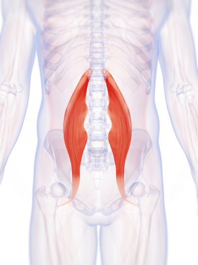
Flexors are flexible muscle tissues that help a person stretch and move. The muscles that help you lift your knee toward your body and bend at the waist are known as the hip flexors. These muscles are located around the upper and inner things and pelvic region. Keeping the hip flexors strong is particularly important for active people and athletes.
Sciatica is a form of radiculopathy caused by compression of the sciatic nerve, the large nerve that travels through the buttocks and extends down the back of the leg. This compression causes shock-like or burning low back pain combined with pain through the buttocks and down one leg, occasionally reaching the foot. In the most extreme cases, when the nerve is pinched between the disc and the adjacent bone, the symptoms may involve not only pain, but numbness and muscle weakness in the leg because of interrupted nerve signaling. The condition may also be caused by a tumor or cyst that presses on the sciatic nerve or its roots.
Athletes are at greater risk of sustaining a lumber spine injury due to physical activity. Whether the sport is skiing, basketball, football, gymnastics, soccer, running, golf, or tennis-the spine undergoes a lot of stress, absorption of pressure, twisting, turning, and even bodily impact. This strenuous activity puts stress on the back that can cause injury to even the finest and most fit athletes.

Bony encroachment: Any condition that results in movement or growth of the vertebrae of the lumbar spine can limit the space (encroachment) for the adjacent spinal cord and nerves. Causes of bony encroachment of the spinal nerves include foraminal narrowing (narrowing of the portal through which the spinal nerve passes from the spinal column, out of the spinal canal to the body, commonly as a result of arthritis), spondylolisthesis (slippage of one vertebra relative to another), and spinal stenosis (compression of the nerve roots or spinal cord by bony spurs or other soft tissues in the spinal canal). Spinal-nerve compression in these conditions can lead to sciatica pain that radiates down the lower extremities. Spinal stenosis can cause lower-extremity pains that worsen with walking and are relieved by resting (mimicking the pains of poor circulation). Treatment of these afflictions varies, depending on their severity, and ranges from rest and exercises to epidural cortisone injections and surgical decompression by removing the bone that is compressing the nervous tissue.

At the start of the 20th century, physicians thought low back pain was caused by inflammation of or damage to the nerves,[99] with neuralgia and neuritis frequently mentioned by them in the medical literature of the time.[100] The popularity of such proposed causes decreased during the 20th century.[100] In the early 20th century, American neurosurgeon Harvey Williams Cushing increased the acceptance of surgical treatments for low back pain.[14] In the 1920s and 1930s, new theories of the cause arose, with physicians proposing a combination of nervous system and psychological disorders such as nerve weakness (neurasthenia) and female hysteria.[99] Muscular rheumatism (now called fibromyalgia) was also cited with increasing frequency.[100]
Whether you lift heavy items for your job or simply have a slipped disk from a pesky athletic injury, lower back pain is likely to plague you at some point in your life. Low back pain can result from an acute injury or from chronic overuse that leads to arthritis. This, in turn, can break down the fluid-filled disks in your spine that act as shock absorbers. Whatever the cause, there are some practices you can do to strengthen your back and keep lower back pain at bay.


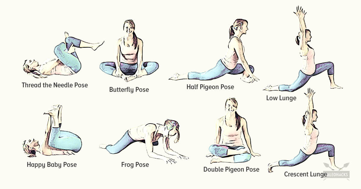
Kneel on your mat with your thighs perpendicular to the floor and tops of your feet facing down. Bring your inner knees together. Slide your feet apart so they are slightly wider than your hips and press the tops of your feet evenly into the mat. Slowly sit down between your feet. Use your hands to turn the top of your thighs inward. Then, lean back onto your forearms and slowly lower torso to floor. Hold for at least 30 seconds.

3. Tendinitis and bursitis Many tendons around the hip connect the muscles to the joint. These tendons can easily become inflamed if you overuse them or participate in strenuous activities. One of the most common causes of tendinitis at the hip joint, especially in runners, is iliotibial band syndrome — the iliotibial band is the thick span of tissue that runs from the outer rim of your pelvis to the outside of your knee.

References to any non-Onnit entity, product, service, person or source of information in this or any other Communication should not be considered an endorsement, either direct or implied, by the host, presenter or distributor of the Communication. The host(s), presenter(s) and/or distributor(s) of this Communication are not responsible for the content of any non-Onnit internet pages referenced in the Communication. Onnit is not liable or responsible for any advice, course of treatment, diagnosis or any other information or services you chose to follow without consulting a qualified medical professional. Before starting any new diet and/or exercise program, always be sure to check with your qualified medical professional.
It’s a common issue, says Prevention advisor Rob Danoff, director of family and emergency medicine residency programs at Aria Health in Philadelphia. "For people who sit a long time at work, the hip flexors and rotators become tight, and the gluteal muscles become weak," he says. "This combination negatively affects our ability to walk, maintain proper posture, and the stability of our spine."
Paget's disease of the bone is a condition of unknown cause in which the bone formation is out of synchrony with normal bone remodeling. This condition results in abnormally weakened bone and deformity and can cause localized bone pain, though it often causes no symptoms. Paget's disease is more common in people over the age of 50. Heredity (genetic background) and certain unusual virus infections have been suggested as causes. Thickening of involved bony areas of the lumbar spine can cause the radiating lower extremity pain of sciatica.
Low back pain that lasts at least one day and limits activity is a common complaint.[7] Globally, about 40% of people have LBP at some point in their lives,[7] with estimates as high as 80% of people in the developed world.[22] Approximately 9 to 12% of people (632 million) have LBP at any given point in time, and nearly one quarter (23.2%) report having it at some point over any one-month period.[7][8] Difficulty most often begins between 20 and 40 years of age.[1] Low back pain is more common among people aged 40–80 years, with the overall number of individuals affected expected to increase as the population ages.[7]

Health Tools Baby Due Date CalculatorBasal Metabolic Rate CalculatorBody Mass Index (BMI) CalculatorCalories Burned CalculatorChild Energy Requirements CalculatorDaily Calcium Requirements CalculatorDaily Fibre Requirements CalculatorIdeal Weight CalculatorInfectious Diseases Exclusion Periods ToolOvulation CalculatorSmoking Cost CalculatorTarget Heart Rate CalculatorWaist-to-hip Ratio Calculator Risk Tests Depression Self-AssessmentErectile Dysfunction ToolMacular Degeneration ToolOsteoporosis Risk TestProstate Symptoms Self-Assessment
In addition to these exercises, there are simple things you can do every day to help reduce your risk of hip flexor pain. If you sit at a desk for long periods of time, try to get up and move around every hour or so. Warm up properly before any physical activity, and stretch regularly at the end of each workout. Your hips will thank you for it!
If you’re worried you’re headed toward a surgeon’s office, there might be hope. According to the Arthritis Foundation, the best way to avoid hip replacement surgery is to get active in an exercise program. In a study, people who participated in an exercise program for 12 weeks were 44 percent less likely to need joint-replacement surgery six years later than those who did not exercise.

Medicine FinderLatest NewsSlip, slap, slop early to save your skinComplementary medicine users have worse cancer survivalVideo: BoredomPut down the tablet; pick up a bookVideo: Lift your mood with some weightsQuicken your step to live longerThis web site is intended for Australian residents and is not a substitute for independent professional advice. Information and interactions contained in this Web site are for information purposes only and are not intended to be used to diagnose, treat, cure or prevent any disease. Further, the accuracy, currency and completeness of the information available on this Web site cannot be guaranteed. Tonic Digital Media Pty Ltd, its affiliates and their respective servants and agents do not accept any liability for any injury, loss or damage incurred by use of or reliance on the information made available via or through myDr whether arising from negligence or otherwise. See Privacy Policy and Disclaimer.2001-2019 myDr.com.au © | All Rights Reserved About UsContact UsDisclaimerPrivacy PolicyAdvertising PolicySitemap

Traction involves the use of weights and pulleys to apply constant or intermittent force to gradually “pull” the skeletal structure into better alignment. Some people experience pain relief while in traction, but that relief is usually temporary. Once traction is released the back pain tends to return. There is no evidence that traction provides any longterm benefits for people with low back pain.
The condition is cauda equina syndrome. It involves “acute loss of function of the neurologic elements (nerve roots) of the spinal canal below the termination (conus) of the spinal cord,” where the nerves spread out like a horse (equina) tail. Again, this condition causes symptoms in the “saddle” of the body: butt, groin, inner thighs. BACK TO TEXT
Discectomy or microdiscectomy may be recommended to remove a disc, in cases where it has herniated and presses on a nerve root or the spinal cord, which may cause intense and enduring pain. Microdiscectomy is similar to a conventional discectomy; however, this procedure involves removing the herniated disc through a much smaller incision in the back and a more rapid recovery. Laminectomy and discectomy are frequently performed together and the combination is one of the more common ways to remove pressure on a nerve root from a herniated disc or bone spur.
The diagnosis of low back pain involves a review of the history of the illness and underlying medical conditions as well as a physical examination. It is essential that a complete story of the back pain be reviewed including injury history, aggravating and alleviating conditions, associated symptoms (fever, numbness, tingling, incontinence, etc.), as well as the duration and progression of symptoms. Aside from routine abdomen and extremity evaluations, rectal and pelvic examinations may eventually be required as well. Further tests for diagnosis of low back pain can be required including blood and urine tests, plain film X-ray tests, CAT scanning, MRI scanning, bone scanning, and tests of the nerves such as electromyograms (EMG) and nerve conduction velocities (NCV).
Why is back pain still a huge problem? Maybe this: “It is extremely difficult to alter the potentially disabling belief among the lay public that low back pain has a structural mechanical cause. An important reason for this is that this belief continues to be regularly reinforced by the conditions of care of a range of ‘hands-on’ providers, for whom idiosyncratic variations of that view are fundamental to their professional existence.”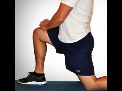
Melissa Conrad Stöppler, MD, is a U.S. board-certified Anatomic Pathologist with subspecialty training in the fields of Experimental and Molecular Pathology. Dr. Stöppler's educational background includes a BA with Highest Distinction from the University of Virginia and an MD from the University of North Carolina. She completed residency training in Anatomic Pathology at Georgetown University followed by subspecialty fellowship training in molecular diagnostics and experimental pathology.
How to: Lie on your back with your right knee bent and foot flat on the floor (a). Extend your left leg up to the ceiling and wrap a strap around the sole of your left foot (b). While holding both ends with your left hand, extend your right arm directly out to the side in order to anchor yourself (c). Slowly let the left leg fall toward the left while keeping your right side grounded. Hold for six to eight breaths, then repeat on the opposite side.

For those with pain localized to the lower back due to disc degeneration, fair evidence supports spinal fusion as equal to intensive physical therapy and slightly better than low-intensity nonsurgical measures.[15] Fusion may be considered for those with low back pain from acquired displaced vertebra that does not improve with conservative treatment,[14] although only a few of those who have spinal fusion experience good results.[15] There are a number of different surgical procedures to achieve fusion, with no clear evidence of one being better than the others.[83] Adding spinal implant devices during fusion increases the risks but provides no added improvement in pain or function.[11]

Imaging is indicated when there are red flags, ongoing neurological symptoms that do not resolve, or ongoing or worsening pain.[5] In particular, early use of imaging (either MRI or CT) is recommended for suspected cancer, infection, or cauda equina syndrome.[5] MRI is slightly better than CT for identifying disc disease; the two technologies are equally useful for diagnosing spinal stenosis.[5] Only a few physical diagnostic tests are helpful.[5] The straight leg raise test is almost always positive in those with disc herniation.[5] Lumbar provocative discography may be useful to identify a specific disc causing pain in those with chronic high levels of low back pain.[41] Similarly, therapeutic procedures such as nerve blocks can be used to determine a specific source of pain.[5] Some evidence supports the use of facet joint injections, transforminal epidural injections and sacroilliac injections as diagnostic tests.[5] Most other physical tests, such as evaluating for scoliosis, muscle weakness or wasting, and impaired reflexes, are of little use.[5]

Radicular pain. This type of pain can occur if a spinal nerve root becomes impinged or inflamed. Radicular pain may follow a nerve root pattern or dermatome down into the buttock and/or leg. Its specific sensation is sharp, electric, burning-type pain and can be associated with numbness or weakness (sciatica). It is typically felt on only one side of the body.