Really a great content. Let me tell you first about hip flexor it is the engine through which our body moves. They control balance, our ability to sit, stand, twist, reach, bend, walk and step. One of my patient also suffering from same problem but due to lack of money he was unable to afford a treatment. So i recommend him a program to unlock hip flexor. If anyone wants they can check it out here ;- https://tinyurl.com/y8yaqs2s Report
Discography may be used when other diagnostic procedures fail to identify the cause of pain. This procedure involves the injection of a contrast dye into a spinal disc thought to be causing low back pain. The fluid’s pressure in the disc will reproduce the person’s symptoms if the disc is the cause. The dye helps to show the damaged areas on CT scans taken following the injection. Discography may provide useful information in cases where people are considering lumbar surgery or when their pain has not responded to conventional treatments.
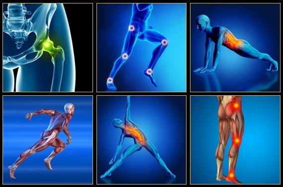
The hip joint is designed to withstand a fair amount of wear and tear, but it’s not indestructible. For example, when you walk, a cushion of cartilage helps prevent friction as the hip bone moves in its socket. With age and use, this cartilage can wear down or become damaged, or the hip bone itself can be fractured during a fall. In fact, more than 300,000 adults over 65 are hospitalized for hip fractures each year, according to the Agency for Healthcare Research and Quality.
For example, your quadriceps muscles are a group of four that are located at the front of the thigh; one of the group members, the rectus femoris flexes the hip, which brings your lower extremity (thigh, lower leg, and foot) forward, in front of you. On the other hand, your hamstring muscles are located at the back of the thigh. When they contract, they extend the lower extremity, bringing it behind you.
In this study, one patient with sciatica was sent for ten MRIs, which produced 49 distinct “findings,” 16 of them unique, none of which occurred in all ten reports. On average, each radiologist made about a dozen errors, seeing one or two things that weren’t there and missing about ten things that were. Yikes. Read a more detailed and informal description of this study.
Electrodiagnostics are procedures that, in the setting of low back pain, are primarily used to confirm whether a person has lumbar radiculopathy. The procedures include electromyography (EMG), nerve conduction studies (NCS), and evoked potential (EP) studies. EMG assesses the electrical activity in a muscle and can detect if muscle weakness results from a problem with the nerves that control the muscles. Very fine needles are inserted in muscles to measure electrical activity transmitted from the brain or spinal cord to a particular area of the body. NCSs are often performed along with EMG to exclude conditions that can mimic radiculopathy. In NCSs, two sets of electrodes are placed on the skin over the muscles. The first set provides a mild shock to stimulate the nerve that runs to a particular muscle. The second set records the nerve’s electrical signals, and from this information nerve damage that slows conduction of the nerve signal can be detected. EP tests also involve two sets of electrodes—one set to stimulate a sensory nerve, and the other placed on the scalp to record the speed of nerve signal transmissions to the brain.
Discography may be used when other diagnostic procedures fail to identify the cause of pain. This procedure involves the injection of a contrast dye into a spinal disc thought to be causing low back pain. The fluid’s pressure in the disc will reproduce the person’s symptoms if the disc is the cause. The dye helps to show the damaged areas on CT scans taken following the injection. Discography may provide useful information in cases where people are considering lumbar surgery or when their pain has not responded to conventional treatments.

Low back pain that lasts at least one day and limits activity is a common complaint.[7] Globally, about 40% of people have LBP at some point in their lives,[7] with estimates as high as 80% of people in the developed world.[22] Approximately 9 to 12% of people (632 million) have LBP at any given point in time, and nearly one quarter (23.2%) report having it at some point over any one-month period.[7][8] Difficulty most often begins between 20 and 40 years of age.[1] Low back pain is more common among people aged 40–80 years, with the overall number of individuals affected expected to increase as the population ages.[7]
Epidural injections of steroid drugs are frequently used to treat sciatica, despite limited evidence for their effectiveness. Moreover, these treatments are based on the assumption that reducing local inflammation in the vertebral column will relieve pain, but an association between structural abnormalities, inflammation, and sciatica symptoms has not been clearly demonstrated. NINDS-funded researchers are using a new imaging technique that can detect inflammation to better understand what causes chronic sciatica pain and to provide evidence to inform treatment selection.
To ease the pain and lower your odds of an injury, don’t try to do too much at once. “Start with just 10 minutes,” says Arina Garg, MD, a rheumatology fellow at The Center for Excellence for Arthritis and Rheumatology at the Louisiana University Health Sciences Center. “Every few days, increase that time by 5 to 10 minutes.” Your goal is to work up to 30 minutes of aerobic exercise, 5 days a week.
If surgery is recommended, neurosurgeons have a variety of options available to help relieve pressure on the nerve roots. If there are several nerve roots and discs causing the pain or if there is degeneration and instability in the spinal column, the neurosurgeon may opt to fuse the vertebrae together with bone grafts and stabilize the vertebrae with instrumentation, including metal plates, screws, rods and cages. A successful fusion will prevent the disc from bulging or herniating again. Following a fusion procedure, a patient may gain restored mobility in the back, including the ability to bend over. He or she will most likely experience more mobility after surgery than before. In addition, the patient may require postoperative physical therapy.
Lie on your back with your knees bent and your feet flat on the floor. Tighten the muscles in your buttocks, then lift your hips off the ground and hold for about five seconds before slowly lowering yourself back down. Be sure to breathe throughout the exercise. As with the first exercise, you can work up to doing 30 repetitions, resting for a few seconds (or longer) between each. “If you start to get tired, stop and rest for a couple of minutes,” Pariser says.

Bone scans are used to detect and monitor infection, fracture, or disorders in the bone. A small amount of radioactive material is injected into the bloodstream and will collect in the bones, particularly in areas with some abnormality. Scanner-generated images can be used to identify specific areas of irregular bone metabolism or abnormal blood flow, as well as to measure levels of joint disease.
If you work at a desk job all day, you might have some areas of your workstation to thank for your back pain. Evaluating your space to make it more ergonomic (back-friendly), can help you experience lower back pain relief and prevent pain from getting worse. Rethinking your workspace for back relief starts with positioning your most important work tools.
I think you should mention that for some people, stretching is not the solution and that it will deteriorate their posture. Some people need stretching, but most people I know need to strengthen their "overstretched" hip flexors. Many people can't do a single hanging leg raise. Check this site if you want to know more about the importance of hip flexors ********** www.smarterpage.wixsite.com/unlock-
Moist heat may help relax your muscles. Put moist heat on the sore area for 10 to 15 minutes at a time before you do warm-up and stretching exercises. Moist heat includes heat patches or moist heating pads that you can buy at most drugstores, a wet washcloth or towel that has been heated in a microwave or the dryer, or a hot shower. Don’t use heat if you have swelling.
Enthoven WT, Geuze J, Scheele J, et al. Prevalence and "Red Flags" Regarding Specified Causes of Back Pain in Older Adults Presenting in General Practice. Phys Ther. 2016 Mar;96(3):305–12. PubMed #26183589. How many cases of back pain in older adults have a serious underlying cause? Only about 6% … but 5% of those are fractures (which are serious, but they aren’t cancer either). The 1% is divided amongst all other serious causes. In this study of 669 patients, a vertebral fracture was found in 33 of them, and the chances of this diagnosis was higher in older patients with more intense pain in the upper back, and (duh) trauma. BACK TO TEXT
Imaging is indicated when there are red flags, ongoing neurological symptoms that do not resolve, or ongoing or worsening pain.[5] In particular, early use of imaging (either MRI or CT) is recommended for suspected cancer, infection, or cauda equina syndrome.[5] MRI is slightly better than CT for identifying disc disease; the two technologies are equally useful for diagnosing spinal stenosis.[5] Only a few physical diagnostic tests are helpful.[5] The straight leg raise test is almost always positive in those with disc herniation.[5] Lumbar provocative discography may be useful to identify a specific disc causing pain in those with chronic high levels of low back pain.[41] Similarly, therapeutic procedures such as nerve blocks can be used to determine a specific source of pain.[5] Some evidence supports the use of facet joint injections, transforminal epidural injections and sacroilliac injections as diagnostic tests.[5] Most other physical tests, such as evaluating for scoliosis, muscle weakness or wasting, and impaired reflexes, are of little use.[5]
So, who cares right? Wrong. Everyone has seen that little old man walking with a cane, hunched over almost to the point of staring at the ground. Do you think he always walked like that? I'd bet you he didn't. Maybe he had an injury that never healed properly, or maybe after spending years and years in a similar position, his body became tighter and tighter until eventually he ended up bent over.
Using heavy weights: Whether you’re a novice weightlifter or a pro, if you suffer from hip pains you should definitely be putting the kettlebell down! Weight-lifting can place too much stress on your body, especially your knees, lower back and hips. You should be focusing instead on gentle strengthening exercises and stretches in order to support your body as it recovers.
The pain of back pain almost always makes it seem worse than it is. The most worrisome causes of back pain rarely cause severe pain, and many common problems (like slipped discs) are usually much less serious than people fear. Only about 1% of back pain is ominous, and even then it’s often still treatable. Most of the 1% are due to cancer, autoimmune disease, or spinal cord damage.
Congenital bone conditions: Congenital causes (existing from birth) of low back pain include scoliosis and spina bifida. Scoliosis is a sideways (lateral) curvature of the spine that can be caused when one lower extremity is shorter than the other (functional scoliosis) or because of an abnormal architecture of the spine (structural scoliosis). Children who are significantly affected by structural scoliosis may require treatment with bracing and/or surgery to the spine. Adults infrequently are treated surgically but often benefit by support bracing. Spina bifida is a birth defect in the bony vertebral arch over the spinal canal, often with absence of the spinous process. This birth defect most commonly affects the lowest lumbar vertebra and the top of the sacrum. Occasionally, there are abnormal tufts of hair on the skin of the involved area. Spina bifida can be a minor bony abnormality without symptoms. However, the condition can also be accompanied by serious nervous abnormalities of the lower extremities.