
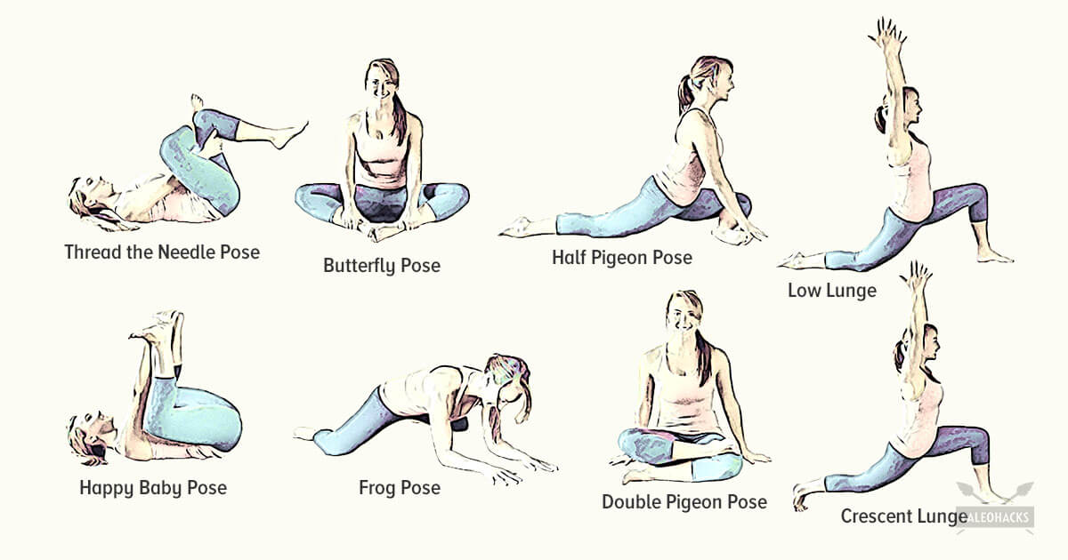
Lie on your back with your knees bent and feet flat on the floor. Place left ankle right below right knee, creating a “four” shape with left leg. Thread left arm through the opening you created with left leg and clasp hands behind right knee. Lift right foot off floor and pull right knee toward chest, flexing left foot. Hold for 30 seconds, then repeat on opposite side.
When it comes to your workouts, low-impact aerobic exercises are generally best and least likely to cause issues, says Kelton Vasileff, M.D., an orthopedic surgeon at Ohio State University Wexner Medical Center. “I recommend swimming, walking, elliptical, cycling, and stationary biking for general exercise,” he says. All of these are great ways to move your body without pounding your joints.
The AANS does not endorse any treatments, procedures, products or physicians referenced in these patient fact sheets. This information is provided as an educational service and is not intended to serve as medical advice. Anyone seeking specific neurosurgical advice or assistance should consult his or her neurosurgeon, or locate one in your area through the AANS’ Find a Board-certified Neurosurgeon” online tool.
Epidural steroid injections are a commonly used short-term option for treating low back pain and sciatica associated with inflammation. Pain relief associated with the injections, however, tends to be temporary and the injections are not advised for long-term use. An NIH-funded randomized controlled trial assessing the benefit of epidural steroid injections for the treatment of chronic low back pain associated with spinal stenosis showed that long-term outcomes were worse among those people who received the injections compared with those who did not.
How to: Sit down with your knees bent and feet flat on the floor in front of you (a). Place your right ankle on top of your left thigh and flex your right foot (b). Put your hands behind your body, fingertips facing away from your body and begin to press your hips toward your heels until you feel a stretch through your outer left hip. Keep your back tall and chest open (c). Hold for six to eight breaths, then repeat on the other side.
Lie on your back with your knees bent and your feet flat on the floor. Tighten the muscles in your buttocks, then lift your hips off the ground and hold for about five seconds before slowly lowering yourself back down. Be sure to breathe throughout the exercise. As with the first exercise, you can work up to doing 30 repetitions, resting for a few seconds (or longer) between each. “If you start to get tired, stop and rest for a couple of minutes,” Pariser says.
Congenital bone conditions: Congenital causes (existing from birth) of low back pain include scoliosis and spina bifida. Scoliosis is a sideways (lateral) curvature of the spine that can be caused when one lower extremity is shorter than the other (functional scoliosis) or because of an abnormal architecture of the spine (structural scoliosis). Children who are significantly affected by structural scoliosis may require treatment with bracing and/or surgery to the spine. Adults infrequently are treated surgically but often benefit by support bracing. Spina bifida is a birth defect in the bony vertebral arch over the spinal canal, often with absence of the spinous process. This birth defect most commonly affects the lowest lumbar vertebra and the top of the sacrum. Occasionally, there are abnormal tufts of hair on the skin of the involved area. Spina bifida can be a minor bony abnormality without symptoms. However, the condition can also be accompanied by serious nervous abnormalities of the lower extremities.
Disc degeneration remains a key cause of chronic low back pain and the pain often persists despite surgery. NIH-funded basic science and preclinical studies are investigating molecular-level mechanisms that cause discs in the spine to degenerate, as well as protective mechanisms involved in disc remodeling that may diminish with advancing age. Such studies may help identify future therapeutic strategies to block degenerative mechanisms or promote remodeling processes. NIH also is funding early research on stem cell approaches to promote disc regeneration and rejuvenate cells of the nucleus pulposus, the jelly-like substance in the center of intervertebral discs that loses water content as people age.
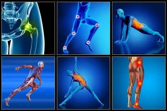
Quick anatomy lesson. When we talk about the hips, we're talking about any muscle that crosses over the hip joint, says Laura Miranda D.P.T., M.S.P.T., C.S.C.S., a New York City-based trainer and creator of the Pursuit training program. Which, there are many, including all of the glute muscles, the hamstrings, the inner thigh muscles, and the psoas muscles (deep core muscles that attach your pelvis to your spine). Each of these muscles has some specific roles, but overall, the hip muscles stabilize your pelvis and thighbone as you move. They also allow you to bend at the hips, lift your legs out to the side (abduct), and bring your legs back in toward one another (adduct). Basically, they do a lot, and when they're weak or tight or otherwise not working in an optimal way, you can not only end up with cranky hips, but other body parts may overcompensate and take on too much work—leaving you with other, seemingly unrelated, issues, like knee pain.
Bleeding in the pelvis is rare without significant trauma and is usually seen in patients who are taking blood-thinning medications, such as warfarin (Coumadin). In these patients, a rapid-onset sciatica pain can be a sign of bleeding in the back of the pelvis and abdomen that is compressing the spinal nerves as they exit to the lower extremities. Infection of the pelvis is infrequent but can be a complication of conditions such as diverticulosis, Crohn's disease, ulcerative colitis, pelvic inflammatory disease with infection of the Fallopian tubes or uterus, and even appendicitis. Pelvic infection is a serious complication of these conditions and is often associated with fever, lowering of blood pressure, and a life-threatening state.

Model Heather Lin grew up in the deep south but is currently hustling in New York, working at a bank. Whether she is biking home from work, deadlifting, kicking a heavy bag, or pouring all of her effort into a bootcamp class, it's important to her to find time in her busy day to work out. She feels her best when she is strong and energized, and blogs about her health and fitness journey at The Herbivore Warrior.
Health Tools Baby Due Date CalculatorBasal Metabolic Rate CalculatorBody Mass Index (BMI) CalculatorCalories Burned CalculatorChild Energy Requirements CalculatorDaily Calcium Requirements CalculatorDaily Fibre Requirements CalculatorIdeal Weight CalculatorInfectious Diseases Exclusion Periods ToolOvulation CalculatorSmoking Cost CalculatorTarget Heart Rate CalculatorWaist-to-hip Ratio Calculator Risk Tests Depression Self-AssessmentErectile Dysfunction ToolMacular Degeneration ToolOsteoporosis Risk TestProstate Symptoms Self-Assessment
Take a step back and think about where you spend most of your day. If you're a young athlete, you probably spend most of your time at school or maybe work or practice and even a little time at home, if you're lucky. Now think about what position your body is in during those periods. I would bet that you spend most of your day sitting down. You may walk to class or run in practice, but the majority of your day is spent in a seated position.
Back pain can be confusing enough as it is. Sometimes it may be difficult deciphering if the pain is even coming from your back, and trying to figure out what causes it can be mind-boggling. And many experts recommend focusing on mechanical sources of your back pain rather than anatomical sources of your pain. Is it a herniated disc, bulging disc, or facet joint arthritis causing your problem? It can be hard to decide, so focusing on what you are doing and the positions you put your back in may be the best way to determine the cause of your back pain.

Even though low back pain can sometimes be treated without major disruption to a person's life, athletes are often reluctant to seek medical help. Many of them deny or minimize complaints in order to avoid consequences, such as: having to decrease activity in order to recover, losing a position or being removed from a team, missing a competition, or letting the team down. Some athletes simply do not want to bother seeing a doctor for pain; they hope it will recover on its own.
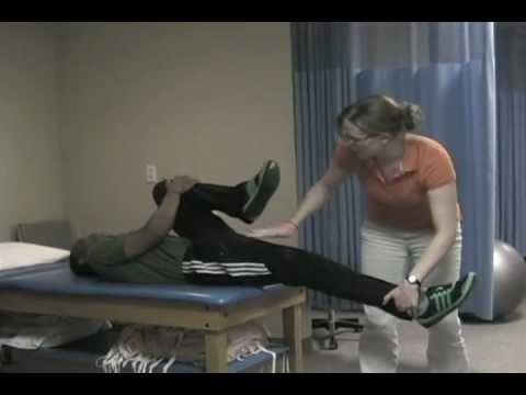
Consider working with a physical therapist to strengthen weak hip muscles. The physical therapists at Beacon Orthopaedists and Sports Medicine can also recommend a sport-specific training program. This is ideal for golfers, swimmers, runners, and any athlete who not only needs to preserve their hip but also strengthen it in order to remain competitive.
Kneel on your mat with your thighs perpendicular to the floor and tops of your feet facing down. Bring your inner knees together. Slide your feet apart so they are slightly wider than your hips and press the tops of your feet evenly into the mat. Slowly sit down between your feet. Use your hands to turn the top of your thighs inward. Then, lean back onto your forearms and slowly lower torso to floor. Hold for at least 30 seconds.

Veritas Health publishes original and accessible health related content written by more than 100 physician authors and peer-reviewed by a 16 member Medical Advisory Board. The Veritas Health platform comprising of Spine-health.com, Arthritis-health.com, Sports-health.com, and Pain-health.com, provides comprehensive information on back pain, arthritis, sports injuries, and chronic pain conditions. For more information visit Veritashealth.com.
Lay on your back on your mat and pull your knees to your chest. Place your hands on the inside arches of your feet and open your knees wider than shoulder-width apart. Keeping your back pressed into the mat as much as possible, press your feet into hands while pulling down on feet, creating resistance. Breathe deeply and hold for at least 30 seconds.

In the majority of cases, a hip sprain begins as a microscopic tear that gradually increases in size with repetitive use of the hip. These types of tears are common in sports like cycling, running, swimming, baseball, and golf due to overuse of the hip. If diagnosed early, Grade I and Grade II strains can be effectively treated with rest and other conservative treatments. Grade III strains, however, are one of the most serious hip injuries. This is especially true if the strain is accompanied by a fracture. If your hip cannot bear weight, it is imperative that you contact an orthopaedist for professional treatment.
Radiculopathy is a condition caused by compression, inflammation and/or injury to a spinal nerve root. Pressure on the nerve root results in pain, numbness, or a tingling sensation that travels or radiates to other areas of the body that are served by that nerve. Radiculopathy may occur when spinal stenosis or a herniated or ruptured disc compresses the nerve root.
Epidural injections of steroid drugs are frequently used to treat sciatica, despite limited evidence for their effectiveness. Moreover, these treatments are based on the assumption that reducing local inflammation in the vertebral column will relieve pain, but an association between structural abnormalities, inflammation, and sciatica symptoms has not been clearly demonstrated. NINDS-funded researchers are using a new imaging technique that can detect inflammation to better understand what causes chronic sciatica pain and to provide evidence to inform treatment selection.

A.D.A.M., Inc. is accredited by URAC, also known as the American Accreditation HealthCare Commission (www.urac.org). URAC's accreditation program is an independent audit to verify that A.D.A.M. follows rigorous standards of quality and accountability. A.D.A.M. is among the first to achieve this important distinction for online health information and services. Learn more about A.D.A.M.'s editorial policy, editorial process and privacy policy. A.D.A.M. is also a founding member of Hi-Ethics and subscribes to the principles of the Health on the Net Foundation (www.hon.ch).
Talmage, J; Belcourt, R; Galper, J; et al. (2011). "Low back disorders". In Kurt T. Hegmann. Occupational medicine practice guidelines : evaluation and management of common health problems and functional recovery in workers (3rd ed.). Elk Grove Village, IL: American College of Occupational and Environmental Medicine. pp. 336, 373, 376–377. ISBN 978-0615452272.
MRI and x-ray for low back pain are surprisingly unreliable,1 because things like bulging discs usually aren’t a deal,2 most back pain goes away on its own,3 and trigger points (“muscle knots”) are common and can be alarmingly intense but aren’t dangerous.4 Most patients are much better off when they feel confident about these things. The power of justified, rational confidence is a huge factor in back pain.5 Sadly, many healthcare professionals continue to perpetuate the idea of fragile backs,6 which undermines that valuable confidence.
Blood tests are not routinely used to diagnose the cause of back pain; however in some cases they may be ordered to look for indications of inflammation, infection, and/or the presence of arthritis. Potential tests include complete blood count, erythrocyte sedimentation rate, and C-reactive protein. Blood tests may also detect HLA-B27, a genetic marker in the blood that is more common in people with ankylosing spondylitis or reactive arthritis (a form of arthritis that occurs following infection in another part of the body, usually the genitourinary tract).
But moving is important for hip and knee OA. It causes your joints to compress and release, bringing blood flow, nutrients, and oxygen into the cartilage. “This can help prolong the function and longevity of your joints,” says Eric Robertson, DPT, a physical therapist and associate professor of clinical physical therapy at the University of Southern California.
Yuri Elkaim is one of the world’s most trusted health and fitness experts. A former pro soccer player turned NYT bestselling author of The All-Day Energy Diet and The All-Day Fat Burning Diet, his clear, science-backed advice has transformed the lives of more than 500,000 men and women and he’s on a mission to help 100 million people by 2040. Read his inspiring story, “From Soccer to Bed to No Hair on My Head” that started it all.
Keeping your hips mobile is important for overall hip function and athletic performance. Mobility refers to the ability of your joints to move through a pain-free, unrestricted range of motion. For cyclists, hip mobility is critical since pedaling occurs in one plane of motion, and after miles and miles in the saddle, hip tightness and restriction may develop. The following movements will help with hip mobility.
This information is not designed to replace a physician's independent judgment about the appropriateness or risks of a procedure for a given patient. Always consult your doctor about your medical conditions or back problem. SpineUniverse does not provide medical advice, diagnosis or treatment. Use of the SpineUniverse.com site is conditional upon your acceptance of our User Agreement

We use cookies and similar technologies to improve your browsing experience, personalize content and offers, show targeted ads, analyze traffic, and better understand you. We may share your information with third-party partners for marketing purposes. To learn more and make choices about data use, visit our Advertising Policy and Privacy Policy. By clicking “Accept and Continue” below, (1) you consent to these activities unless and until you withdraw your consent using our rights request form, and (2) you consent to allow your data to be transferred, processed, and stored in the United States.
If you are experiencing true numbness14 around the groin and buttocks and/or failure of bladder or bowel control, please consider it a serious emergency — do not wait to see if it goes away. These symptoms indicate spinal cord injury or compression15 and require immediate medical attention. (Few people will have symptoms like this without having already decided it’s an emergency, but I have to cover all the bases here.)
^ Coxib and traditional NSAID Trialists' (CNT) Collaboration, Bhala N, Emberson J, Merhi A, Abramson S, Arber N, Baron JA, Bombardier C, Cannon C, Farkouh ME, FitzGerald GA, Goss P, Halls H, Hawk E, Hawkey C, Hennekens C, Hochberg M, Holland LE, Kearney PM, Laine L, Lanas A, Lance P, Laupacis A, Oates J, Patrono C, Schnitzer TJ, Solomon S, Tugwell P, Wilson K, Wittes J, Baigent C (Aug 31, 2013). "Vascular and upper gastrointestinal effects of non-steroidal anti-inflammatory drugs: meta-analyses of individual participant data from randomised trials". Lancet. 382 (9894): 769–79. doi:10.1016/S0140-6736(13)60900-9. PMC 3778977. PMID 23726390.
In this study, one patient with sciatica was sent for ten MRIs, which produced 49 distinct “findings,” 16 of them unique, none of which occurred in all ten reports. On average, each radiologist made about a dozen errors, seeing one or two things that weren’t there and missing about ten things that were. Yikes. Read a more detailed and informal description of this study.
Discography may be used when other diagnostic procedures fail to identify the cause of pain. This procedure involves the injection of a contrast dye into a spinal disc thought to be causing low back pain. The fluid’s pressure in the disc will reproduce the person’s symptoms if the disc is the cause. The dye helps to show the damaged areas on CT scans taken following the injection. Discography may provide useful information in cases where people are considering lumbar surgery or when their pain has not responded to conventional treatments.

