
^ Coxib and traditional NSAID Trialists' (CNT) Collaboration, Bhala N, Emberson J, Merhi A, Abramson S, Arber N, Baron JA, Bombardier C, Cannon C, Farkouh ME, FitzGerald GA, Goss P, Halls H, Hawk E, Hawkey C, Hennekens C, Hochberg M, Holland LE, Kearney PM, Laine L, Lanas A, Lance P, Laupacis A, Oates J, Patrono C, Schnitzer TJ, Solomon S, Tugwell P, Wilson K, Wittes J, Baigent C (Aug 31, 2013). "Vascular and upper gastrointestinal effects of non-steroidal anti-inflammatory drugs: meta-analyses of individual participant data from randomised trials". Lancet. 382 (9894): 769–79. doi:10.1016/S0140-6736(13)60900-9. PMC 3778977. PMID 23726390.
Or anything else. Pain is a poor indicator, period! The human nervous system is really terrible about this: it routinely produces false alarms, and alarms that are much too loud. See Pain is Weird: Pain science reveals a volatile, misleading sensation that is often more than just a symptom, and sometimes worse than whatever started it. BACK TO TEXT

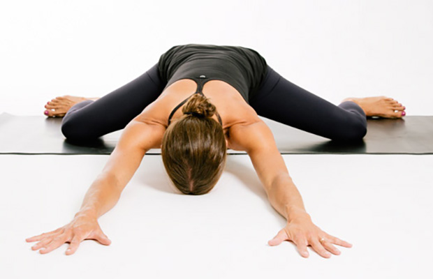
Following any period of prolonged inactivity, a regimen of low-impact exercises is advised. Speed walking, swimming, or stationary bike riding 30 minutes daily can increase muscle strength and flexibility. Yoga also can help stretch and strengthen muscles and improve posture. Consult a physician for a list of low-impact, age-appropriate exercises that are specifically targeted to strengthening lower back and abdominal muscles.
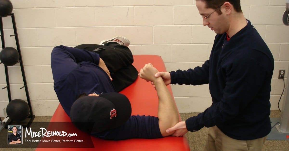
Electrodiagnostics are procedures that, in the setting of low back pain, are primarily used to confirm whether a person has lumbar radiculopathy. The procedures include electromyography (EMG), nerve conduction studies (NCS), and evoked potential (EP) studies. EMG assesses the electrical activity in a muscle and can detect if muscle weakness results from a problem with the nerves that control the muscles. Very fine needles are inserted in muscles to measure electrical activity transmitted from the brain or spinal cord to a particular area of the body. NCSs are often performed along with EMG to exclude conditions that can mimic radiculopathy. In NCSs, two sets of electrodes are placed on the skin over the muscles. The first set provides a mild shock to stimulate the nerve that runs to a particular muscle. The second set records the nerve’s electrical signals, and from this information nerve damage that slows conduction of the nerve signal can be detected. EP tests also involve two sets of electrodes—one set to stimulate a sensory nerve, and the other placed on the scalp to record the speed of nerve signal transmissions to the brain.

Overall, the outcome for acute low back pain is positive. Pain and disability usually improve a great deal in the first six weeks, with complete recovery reported by 40 to 90%.[2] In those who still have symptoms after six weeks, improvement is generally slower with only small gains up to one year. At one year, pain and disability levels are low to minimal in most people. Distress, previous low back pain, and job satisfaction are predictors of long-term outcome after an episode of acute pain.[2] Certain psychological problems such as depression, or unhappiness due to loss of employment may prolong the episode of low back pain.[13] Following a first episode of back pain, recurrences occur in more than half of people.[23]
As with any sort of pain, it's crucial to figure out the source so you can properly treat it. Sharp or stabbing pain that extends beyond your low back or is accompanied by symptoms like abdominal pain, nausea, and vomiting, could be signs of various other conditions and definitely warrant a trip to the doctor. If you have a history of lower back injuries or disc problems, always see your doctor before trying any new exercise.
Magnetic resonance imaging (MRI) uses a magnetic force instead of radiation to create a computer-generated image. Unlike x-ray, which shows only bony structures, MRI scans also produce images of soft tissues such as muscles, ligaments, tendons, and blood vessels. An MRI may be ordered if a problem such as infection, tumor, inflammation, disc herniation or rupture, or pressure on a nerve is suspected. MRI is a noninvasive way to identify a condition requiring prompt surgical treatment. However, in most instances, unless there are “red flags” in the history or physical exam, an MRI scan is not necessary during the early phases of low back pain.
The hip joint is designed to withstand a fair amount of wear and tear, but it’s not indestructible. For example, when you walk, a cushion of cartilage helps prevent friction as the hip bone moves in its socket. With age and use, this cartilage can wear down or become damaged, or the hip bone itself can be fractured during a fall. In fact, more than 300,000 adults over 65 are hospitalized for hip fractures each year, according to the Agency for Healthcare Research and Quality.
Stretching is your next move, but not just any stretches. “Before your workout, you want to go for dynamic stretches, or stretches that put the joint through a full range of motion,” says Lefkowith. Moves like squats and lunges will get your muscles fired up (especially if you focus on squeezing your butt at the top of those squats), says Lefkowith.
This Australian study concluded that “prognosis is moderately optimistic for patients with chronic low back pain,” contradicting the common fear that any low back pain that lasts longer than 6-9 weeks will become a long-term chronic problem. This evidence is the first of its kind, a rarity in low back pain research, a field where almost everything has been studied to death. “Many studies provide good evidence for the prognosis of acute low back pain,” the authors explain. “Relatively few provide good evidence for the prognosis of chronic low back pain.”
Most low back pain is acute, or short term, and lasts a few days to a few weeks. It tends to resolve on its own with self-care and there is no residual loss of function. The majority of acute low back pain is mechanical in nature, meaning that there is a disruption in the way the components of the back (the spine, muscle, intervertebral discs, and nerves) fit together and move.
If your hips are killing you, you probably spend a lot of time sitting – in the car, at work, on that spinning seat – which puts your hips in near-constant “flexion”, says Cori Lefkowith, NASM-certified personal trainer and owner of Redefining Strength in Orange County, California. Even running involves a repetitive flexion movement that can cause pain.
The use of lumbar supports in the form of wide elastic bands that can be tightened to provide support to the lower back and abdominal muscles to prevent low back pain remains controversial. Such supports are widely used despite a lack of evidence showing that they actually prevent pain. Multiple studies have determined that the use of lumbar supports provides no benefit in terms of the prevention and treatment of back pain. Although there have been anecdotal case reports of injury reduction among workers using lumbar support belts, many companies that have back belt programs also have training and ergonomic awareness programs. The reported injury reduction may be related to a combination of these or other factors. Furthermore, some caution is advised given that wearing supportive belts may actually lead to or aggravate back pain by causing back muscles to weaken from lack of use.
In this study, one patient with sciatica was sent for ten MRIs, which produced 49 distinct “findings,” 16 of them unique, none of which occurred in all ten reports. On average, each radiologist made about a dozen errors, seeing one or two things that weren’t there and missing about ten things that were. Yikes. Read a more detailed and informal description of this study.
^ Jump up to: a b c d e f Qaseem, A; Wilt, TJ; McLean, RM; Forciea, MA; Clinical Guidelines Committee of the American College of, Physicians. (4 April 2017). "Noninvasive Treatments for Acute, Subacute, and Chronic Low Back Pain: A Clinical Practice Guideline From the American College of Physicians". Annals of Internal Medicine. 166 (7): 514–530. doi:10.7326/M16-2367. PMID 28192789.
Take a step back and think about where you spend most of your day. If you're a young athlete, you probably spend most of your time at school or maybe work or practice and even a little time at home, if you're lucky. Now think about what position your body is in during those periods. I would bet that you spend most of your day sitting down. You may walk to class or run in practice, but the majority of your day is spent in a seated position.

