Osteoarthritis. This condition results from wear and tear of the disc and facet joints. It causes pain, inflammation, instability, and stenosis to a variable degree, and can occur at a single level or multiple levels of the lower spine. Spinal osteoarthritis is associated with aging and is slowly progressive. It is also referred to as spondylosis or degenerative joint disease.
If you have hip arthritis, work on building up the muscles in your outer thigh for added support. Lie on your pain-free side and lift the leg with arthritis up about six inches, hold for two or three seconds, and lower it again, Humphrey says. Start with one set of 10 repetitions and build up to three sets. Repeat on the other side unless it is too painful. This exercise can aggravate your symptoms if you have hip pain from bursitis.

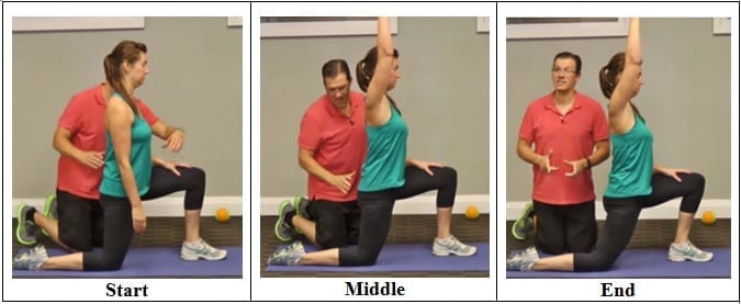
Stand tall with your hips square and bend your right knee, bringing your foot towards your bum. Grab the right foot with your right hand and actively pull the foot closer to your glutes. As you do this, send the right knee down towards the ground and keep both knees together. squeeze your butt to promote a posterior pelvic tilt and hold — then switch sides.
Quick anatomy lesson. When we talk about the hips, we're talking about any muscle that crosses over the hip joint, says Laura Miranda D.P.T., M.S.P.T., C.S.C.S., a New York City-based trainer and creator of the Pursuit training program. Which, there are many, including all of the glute muscles, the hamstrings, the inner thigh muscles, and the psoas muscles (deep core muscles that attach your pelvis to your spine). Each of these muscles has some specific roles, but overall, the hip muscles stabilize your pelvis and thighbone as you move. They also allow you to bend at the hips, lift your legs out to the side (abduct), and bring your legs back in toward one another (adduct). Basically, they do a lot, and when they're weak or tight or otherwise not working in an optimal way, you can not only end up with cranky hips, but other body parts may overcompensate and take on too much work—leaving you with other, seemingly unrelated, issues, like knee pain.
Dr. Shiel received a Bachelor of Science degree with honors from the University of Notre Dame. There he was involved in research in radiation biology and received the Huisking Scholarship. After graduating from St. Louis University School of Medicine, he completed his Internal Medicine residency and Rheumatology fellowship at the University of California, Irvine. He is board-certified in Internal Medicine and Rheumatology.
Bony encroachment: Any condition that results in movement or growth of the vertebrae of the lumbar spine can limit the space (encroachment) for the adjacent spinal cord and nerves. Causes of bony encroachment of the spinal nerves include foraminal narrowing (narrowing of the portal through which the spinal nerve passes from the spinal column, out of the spinal canal to the body, commonly as a result of arthritis), spondylolisthesis (slippage of one vertebra relative to another), and spinal stenosis (compression of the nerve roots or spinal cord by bony spurs or other soft tissues in the spinal canal). Spinal-nerve compression in these conditions can lead to sciatica pain that radiates down the lower extremities. Spinal stenosis can cause lower-extremity pains that worsen with walking and are relieved by resting (mimicking the pains of poor circulation). Treatment of these afflictions varies, depending on their severity, and ranges from rest and exercises to epidural cortisone injections and surgical decompression by removing the bone that is compressing the nervous tissue.
Infection of the discs (septic discitis) and bone (osteomyelitis) is extremely rare. These conditions lead to localized pain associated with fever. The bacteria found when these tissues are tested with laboratory cultures include Staphylococcus aureus and Mycobacterium tuberculosis (TB bacteria). TB infection in the spine is called Pott's disease. These are each very serious conditions requiring long courses of antibiotics. The sacroiliac joints rarely become infected with bacteria. Brucellosis is a bacterial infection that can involve the sacroiliac joints and is usually transmitted in raw goat's milk.
Approximately 15 degrees of hip extension is required to walk normally. If hip flexors are tight then in order to walk, compensatory movement needs to take place through the lower back causing back pain and premature disc degeneration. Like other joints, if we fail to take them through their full range on a regular basis we eventually lose mobility.
But moving is important for hip and knee OA. It causes your joints to compress and release, bringing blood flow, nutrients, and oxygen into the cartilage. “This can help prolong the function and longevity of your joints,” says Eric Robertson, DPT, a physical therapist and associate professor of clinical physical therapy at the University of Southern California.

MRI and x-ray for low back pain are surprisingly unreliable,1 because things like bulging discs usually aren’t a deal,2 most back pain goes away on its own,3 and trigger points (“muscle knots”) are common and can be alarmingly intense but aren’t dangerous.4 Most patients are much better off when they feel confident about these things. The power of justified, rational confidence is a huge factor in back pain.5 Sadly, many healthcare professionals continue to perpetuate the idea of fragile backs,6 which undermines that valuable confidence.

We implement a variety of security measures to maintain the safety of your personal information when you place an order or enter, submit, or access any information on our website. We incorporate physical, electronic, and administrative procedures to safeguard the confidentiality of your personal information, including Secure Sockets Layer (SSL) for the encryption of all financial transactions through the website. We use industry-standard, 256bit SSL encryption to protect your personal information online, and we also take several steps to protect your personal information in our facilities. For example, when you visit the website, you access servers that are kept in a secure physical environment, behind a locked cage and a hardware firewall. After a transaction, your credit card information is not stored on our servers.

Imaging is indicated when there are red flags, ongoing neurological symptoms that do not resolve, or ongoing or worsening pain.[5] In particular, early use of imaging (either MRI or CT) is recommended for suspected cancer, infection, or cauda equina syndrome.[5] MRI is slightly better than CT for identifying disc disease; the two technologies are equally useful for diagnosing spinal stenosis.[5] Only a few physical diagnostic tests are helpful.[5] The straight leg raise test is almost always positive in those with disc herniation.[5] Lumbar provocative discography may be useful to identify a specific disc causing pain in those with chronic high levels of low back pain.[41] Similarly, therapeutic procedures such as nerve blocks can be used to determine a specific source of pain.[5] Some evidence supports the use of facet joint injections, transforminal epidural injections and sacroilliac injections as diagnostic tests.[5] Most other physical tests, such as evaluating for scoliosis, muscle weakness or wasting, and impaired reflexes, are of little use.[5]

The problem is that these muscles aren't designed to be prime movers—they're designed to support the action of the glutes. Inability of activating the glutes can result in low back pain (low back muscles compensating), hamstring strains (overacting hamstrings), hip pain (resulting from hamstring-dominant hip extension) and knee pain (poor glute medius strength).
The management of low back pain often includes medications for the duration that they are beneficial. With the first episode of low back pain the hope is a complete cure; however, if the problem becomes chronic, the goals may change to pain management and the recovery of as much function as possible. As pain medications are only somewhat effective, expectations regarding their benefit may differ from reality, and this can lead to decreased satisfaction.[13]
When you're working to strengthen the core, you'll want to focus on exercises that don't exacerbate lower back issues. "It's important to find out which movements (flexion, extension, rotation) cause pain or discomfort and to avoid those movements, while continuing to work into ranges that are not provoking," Dircksen says. Crockford suggests focusing on exercises that keep the core stable and avoiding twisting movements to avoid exacerbating pain.
5. Feel free as a bird. Open up those hips with yoga’s pigeon pose! Start on all fours with hands below the shoulders and knees below the hips. Bring the right knee forward until it touches the right hand and place the leg flat on the ground across the body (the right foot is now on the left side of the body, parallel to the front of the mat). Drop left leg to the ground, and extend it back with toes turned under. Keep the hips level, inhale, and walk hands forward. Exhale, and fold the torso over, lowering elbows to the floor. Stay in this position for 5-10 breaths before coming back up to switch sides.