
The presence of certain signs, termed red flags, indicate the need for further testing to look for more serious underlying problems, which may require immediate or specific treatment.[5][36] The presence of a red flag does not mean that there is a significant problem. It is only suggestive,[37][38] and most people with red flags have no serious underlying problem.[3][1] If no red flags are present, performing diagnostic imaging or laboratory testing in the first four weeks after the start of the symptoms has not been shown to be useful.[5]

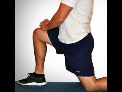
How to: Sit on the floor with knees bent so that your right shin is positioned in front of you, your left shin behind you and your left hip dropped all of the way to the floor (a). Inhale and press your left hip forward until you feel a stretch in the front of your hip (b). Exhale and press left hip back to the floor. That’s one rep (c). Complete six to eight reps, working each time to increase your range of motion. Repeat on the opposite side.
The hip is a very stable ball and socket type joint with an inherently large range of motion. The hip contains some of the largest muscle in the body as well as some of the smallest. Most people lack mobility due to a relatively sedentary lifestyle. Periods of prolonged sitting results in tightness of the hip flexors and hamstrings. Tightness in the muscles and ligaments can created joint forces that result in arthritis, postural problems, bursitis, and mechanical back pain.
Our Keep-It™ guarantee is valid for the first-time purchase of a formula, and redeemable up to three months (90 days) after the purchase date. Multiple bottles, foods, apparel and gear do not fall under this guarantee, however, they may be applicable for return. Fitness equipment, personal care products, knowledge purchases, and DVDs are not eligible for return or refund. For more information and a full list of products that qualify, visit our Keep-It™ page. Further details can be found on our Refund Policy support page.
There are a few most common causes of hip pain. The first thing to distinguish is to identify which pain is coming from the hip, as opposed to some other source. So there are four causes of hip pain, and the pain can come from muscles, ligaments, tendons, and within the joint itself. But those types of pain present in different ways. So those are the most important distinguishing factors to find out if the hip actually is the cause of the pain.
Why is back pain still a huge problem? Maybe this: “It is extremely difficult to alter the potentially disabling belief among the lay public that low back pain has a structural mechanical cause. An important reason for this is that this belief continues to be regularly reinforced by the conditions of care of a range of ‘hands-on’ providers, for whom idiosyncratic variations of that view are fundamental to their professional existence.”
^ Paige, Neil M.; Miake-Lye, Isomi M.; Booth, Marika Suttorp; Beroes, Jessica M.; Mardian, Aram S.; Dougherty, Paul; Branson, Richard; Tang, Baron; Morton, Sally C.; Shekelle, Paul G. (11 April 2017). "Association of Spinal Manipulative Therapy With Clinical Benefit and Harm for Acute Low Back Pain". JAMA. 317 (14): 1451–1460. doi:10.1001/jama.2017.3086. PMC 5470352. PMID 28399251.
The diagnosis of low back pain involves a review of the history of the illness and underlying medical conditions as well as a physical examination. It is essential that a complete story of the back pain be reviewed including injury history, aggravating and alleviating conditions, associated symptoms (fever, numbness, tingling, incontinence, etc.), as well as the duration and progression of symptoms. Aside from routine abdomen and extremity evaluations, rectal and pelvic examinations may eventually be required as well. Further tests for diagnosis of low back pain can be required including blood and urine tests, plain film X-ray tests, CAT scanning, MRI scanning, bone scanning, and tests of the nerves such as electromyograms (EMG) and nerve conduction velocities (NCV).
Pain on the outside of the hip is most commonly due to greater trochanteric bursitis. The greater trochanter is the protrusion where the thigh bone juts outward at the base of the neck (which connects the ball to the femur and is the site of hip stress fractures). A lubricating sac (or bursa) lies over the boney protrusion so that the surrounding muscles do not rub directly on the bone. The top region of the iliotibial (IT) band, known as the tensor fascia lata, is commonly involved in greater trochanteric bursitis.
In this study, one patient with sciatica was sent for ten MRIs, which produced 49 distinct “findings,” 16 of them unique, none of which occurred in all ten reports. On average, each radiologist made about a dozen errors, seeing one or two things that weren’t there and missing about ten things that were. Yikes. Read a more detailed and informal description of this study.
Stop listening to other people’s horror stories. You know the scenario: You are bent over in obvious pain, waiting to see the doctor, and the person next to you tells you a 10-minute tale of how their Uncle Gordon had low back pain that required injections and surgery. But the pain still didn’t go away. Stop listening to these terrible stories. Most low back pain is short-lived and can be managed quite effectively with exercise and postural correction. Of course, some low back conditions are serious and require surgery, but that is a conversation you should have with your doctor, not the guy in the waiting room.

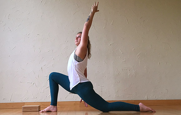
Brace your core and bend your knees to lower down into a split squat. Your left knee should ideally form a 90-degree angle so that your thigh is parallel to the ground, and your right knee is hovering above the floor. (Quick position check: your left foot should be stepped out far enough that you can do this without letting your left knee go past your left toes—if you can't, hop your left foot out a bit farther away from the bench.)
NINDS health-related material is provided for information purposes only and does not necessarily represent endorsement by or an official position of the National Institute of Neurological Disorders and Stroke or any other Federal agency. Advice on the treatment or care of an individual patient should be obtained through consultation with a physician who has examined that patient or is familiar with that patient's medical history.
Hamstring squeeze. Use the machine that works your hamstrings; you will either lie on your stomach or sit with a pad behind your knee. Push against the pad, moving your knee up toward the ceiling or backward (depending on which position you’re in). “In other words, bend your knees,” Pariser says. But to avoid cramps in your hamstring muscles, don’t bend your knee so much that your heels are too close to your buttocks.
The multifidus muscles run up and down along the back of the spine, and are important for keeping the spine straight and stable during many common movements such as sitting, walking and lifting.[12] A problem with these muscles is often found in someone with chronic low back pain, because the back pain causes the person to use the back muscles improperly in trying to avoid the pain.[31] The problem with the multifidus muscles continues even after the pain goes away, and is probably an important reason why the pain comes back.[31] Teaching people with chronic low back pain how to use these muscles is recommended as part of a recovery program.[31]
In both younger and older patients, vertebral fractures take weeks to heal with rest and pain relievers. Compression fractures of vertebrae associated with osteoporosis can also be treated with a procedure called vertebroplasty or kyphoplasty, which can help to reduce pain. In this procedure, a balloon is inflated in the compressed vertebra, often returning some of its lost height. Subsequently, a "cement" (methymethacrylate) is injected into the balloon and remains to retain the structure and height of the body of the vertebra. Pain is relieved as the height of the collapsed vertebra is restored.

