
If you have hip pain, you may benefit from the skilled services of a physical therapist to help determine the cause of your pain. Your PT can work with you to develop a treatment strategy to treat your hip pain or hip discomfort. Understanding why your hip is hurting can help your physical therapist and doctor prescribe the right treatment regimen for your specific condition.

The problem is that these muscles aren't designed to be prime movers—they're designed to support the action of the glutes. Inability of activating the glutes can result in low back pain (low back muscles compensating), hamstring strains (overacting hamstrings), hip pain (resulting from hamstring-dominant hip extension) and knee pain (poor glute medius strength).
2016 — More editing, more! Added some better information about pain being a poor indicator, and the role of myofascial trigger points. This article has become extremely busy in the last couple months — about 4,000 readers per day, as described here — so I am really polishing it and making sure that it’s the best possible answer to people’s fears about back pain.

Talmage, J; Belcourt, R; Galper, J; et al. (2011). "Low back disorders". In Kurt T. Hegmann. Occupational medicine practice guidelines : evaluation and management of common health problems and functional recovery in workers (3rd ed.). Elk Grove Village, IL: American College of Occupational and Environmental Medicine. pp. 336, 373, 376–377. ISBN 978-0615452272.

Endometriosis (when the uterus lining grows somewhere else) can cause pelvic tenderness, which some women describe as hip pain. Pain from the back and spine also can be felt around the buttocks and hip, Siegrist says. Sciatica, a pinched nerve, typically affects one side of the body and can cause pain in the back of the right or left hip — the pain from sciatica can start in your lower back and travel down to your buttocks and legs.

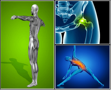
Work on strengthening all of your core muscles and glutes. These muscles work together to give you balance and stability and to help you move through the activities involved in daily living, as well as exercise and sports. When one set of these muscles is weak or tight, it can cause injury or pain in another, so make sure you pay equal attention to all of them.
For example, your quadriceps muscles are a group of four that are located at the front of the thigh; one of the group members, the rectus femoris flexes the hip, which brings your lower extremity (thigh, lower leg, and foot) forward, in front of you. On the other hand, your hamstring muscles are located at the back of the thigh. When they contract, they extend the lower extremity, bringing it behind you. 
Strength training is another key part of the “do” category, Dr. Vasileff says. “It’s a good idea to focus on quad, hamstring, and glute strength,” he says. These muscles surround your hips and provide support, along with your core—which is another area to focus on. “Strengthening your core helps to normalize your walking pattern and stabilize how your pelvis and hips move,” Dr. Vasileff says. That translates to less pain and better hip mobility.
Beacon Orthopaedics & Sports Medicine, Ltd. complies with applicable Federal civil rights laws and does not discriminate on the basis of race, color, national origin, age, disability, or sex. If you speak a language other than English, language assistance services, free of charge, are available to you. Call the Call Center at (513) 354-3700. Español (Spanish): ATENCIÓN: Si habla español, tiene a su disposición servicios gratuitos de asistencia lingüística. Llame al Call Center at (513) 354-3700. 繁體中文 (Chinese): 注意:如果您使用繁體中文,您可以免費獲得語言援助服務。請致電 Call Center at (513) 354-3700.
Located deep in the front of the hip and connecting the leg, pelvis, and abdomen, the hip flexors— surprise, surprise— flex the hip. But despite being some of the most powerful muscles in our bodies (with a clearly important role), it’s easy to neglect our poor hip flexors— often without even knowing it. It turns out just working at a desk all day (guilty!) can really weaken hip flexors since they tend to shorten up while in a seated position. This tightness disrupts good posture and is a common cause of lower back pain. Weakened hip flexors can also increase the risk of foot, ankle, and knee injuries (especially among runners) Hip muscle weakness and overuse injuries in recreational runners. Niemuth, P.E., Johnson, R.J., Myers, M.J., et al. Rocky Mountain University of Health Professions, Provo, VT. Clinical Journal of Sport Medicine, 2005 Jan; 15 (1): 14-21.. So be sure to get up, stand up every hour or so! And giving the hip flexors some extra attention is not just about injury prevention. Adding power to workouts, working toward greater flexibility, and getting speedier while running is also, as they say, all in the hips The effect of walking speed on muscle function and mechanical energetics. Neptune, R.R., Sasaki, K., and Kautz, S.A. Department of Mechanical Engineering, The University of Texas, Austin, TX. Gait & Posture, 2008 Jul; 28 (1): 135-43..
The iliopsoas muscles are a group of two muscles—the psoas muscle and the iliacus muscle—located toward the front of the inner hip. The psoas muscles, in particular, is located in the lumbar (lower) region of the spine and extends through the pelvis to the femur. The iliopsoas muscles are the primary hip flexors, pulling the knee up off the ground when it contracts. Because the psoas muscle is also connected to the spine, it contributes to upright posture, assists in lumbar spine movement, and influences the spine’s curve.
Discography may be used when other diagnostic procedures fail to identify the cause of pain. This procedure involves the injection of a contrast dye into a spinal disc thought to be causing low back pain. The fluid’s pressure in the disc will reproduce the person’s symptoms if the disc is the cause. The dye helps to show the damaged areas on CT scans taken following the injection. Discography may provide useful information in cases where people are considering lumbar surgery or when their pain has not responded to conventional treatments.
Recovering from a hip flexor injury can take time, but proper supervision and guidance from a musculoskeletal professional can minimize downtime and pain. Additionally, there are a few stretches you can do at home to recover from a hip flexor injury. The following stretch increases hip flexor dexterity, while helping alleviate pain in and around the hip and upper thigh.
Stop listening to other people’s horror stories. You know the scenario: You are bent over in obvious pain, waiting to see the doctor, and the person next to you tells you a 10-minute tale of how their Uncle Gordon had low back pain that required injections and surgery. But the pain still didn’t go away. Stop listening to these terrible stories. Most low back pain is short-lived and can be managed quite effectively with exercise and postural correction. Of course, some low back conditions are serious and require surgery, but that is a conversation you should have with your doctor, not the guy in the waiting room.

For the 31 million Americans who suffer from daily back pain, relief can be hard to find. In fact, back pain is the single leading cause of disability worldwide and the second most common reason for visits to the doctor. While it can be caused by many different things—extended periods of sitting or standing at work, bad posture, stress, an overly-strenuous workout or helping a friend move, to mention a few—once back pain hits, it can stick around for a long time. Your gut instinct might be to stay frozen until it goes away, but the best thing for your back is to keep it moving with gentle stretches. In fact, a regular routine of a few quick exercises can help you reduce your back pain without a trip to the doctor. Try to do the following exercises every morning and again at night.
That is, the parts of your body that touch a saddle when riding a horse: groin, buttock, and inner thighs. I experienced rather intense, terrifying awareness of symptoms in this area in the aftermath of my wife’s car accident in early 2010. With a mangled T12 vertebrae, she was at real risk of exactly this problem. Fortunately, she escaped that quite serious problem. But, sheesh, I was vigilant about it for a while! “Honey, any numbness in your saddle area today?” BACK TO TEXT
The AANS does not endorse any treatments, procedures, products or physicians referenced in these patient fact sheets. This information is provided as an educational service and is not intended to serve as medical advice. Anyone seeking specific neurosurgical advice or assistance should consult his or her neurosurgeon, or locate one in your area through the AANS’ Find a Board-certified Neurosurgeon” online tool.
Bone scans are used to detect and monitor infection, fracture, or disorders in the bone. A small amount of radioactive material is injected into the bloodstream and will collect in the bones, particularly in areas with some abnormality. Scanner-generated images can be used to identify specific areas of irregular bone metabolism or abnormal blood flow, as well as to measure levels of joint disease.

Parts of the pain sensation and processing system may not function properly; creating the feeling of pain when no outside cause exists, signaling too much pain from a particular cause, or signaling pain from a normally non-painful event. Additionally, the pain modulation mechanisms may not function properly. These phenomena are involved in chronic pain.[12] 
Exercise therapy is effective in decreasing pain and improving function for those with chronic low back pain.[50] It also appears to reduce recurrence rates for as long as six months after the completion of program[61] and improves long-term function.[57] There is no evidence that one particular type of exercise therapy is more effective than another.[62] The Alexander technique appears useful for chronic back pain,[63] and there is tentative evidence to support the use of yoga.[64] Transcutaneous electrical nerve stimulation (TENS) has not been found to be effective in chronic low back pain.[65] Evidence for the use of shoe insoles as a treatment is inconclusive.[51] Peripheral nerve stimulation, a minimally-invasive procedure, may be useful in cases of chronic low back pain that do not respond to other measures, although the evidence supporting it is not conclusive, and it is not effective for pain that radiates into the leg.[66]
In addition to strengthening the core muscles, it's also important to address any mobility problems, says Jacque Crockford, M.S., C.S.C.S., exercise physiology content manager at American Council on Exercise, which can sometimes be what's causing pain. If specific movements like twisting or bending or extending your spine feel uncomfortable, there may be mobility (flexibility) issues at play. Doing some gentle stretching (like these yoga poses) might help. (If it gets worse with those stretches, stop and see a doctor.)
Electrodiagnostics are procedures that, in the setting of low back pain, are primarily used to confirm whether a person has lumbar radiculopathy. The procedures include electromyography (EMG), nerve conduction studies (NCS), and evoked potential (EP) studies. EMG assesses the electrical activity in a muscle and can detect if muscle weakness results from a problem with the nerves that control the muscles. Very fine needles are inserted in muscles to measure electrical activity transmitted from the brain or spinal cord to a particular area of the body. NCSs are often performed along with EMG to exclude conditions that can mimic radiculopathy. In NCSs, two sets of electrodes are placed on the skin over the muscles. The first set provides a mild shock to stimulate the nerve that runs to a particular muscle. The second set records the nerve’s electrical signals, and from this information nerve damage that slows conduction of the nerve signal can be detected. EP tests also involve two sets of electrodes—one set to stimulate a sensory nerve, and the other placed on the scalp to record the speed of nerve signal transmissions to the brain.
Imaging is indicated when there are red flags, ongoing neurological symptoms that do not resolve, or ongoing or worsening pain.[5] In particular, early use of imaging (either MRI or CT) is recommended for suspected cancer, infection, or cauda equina syndrome.[5] MRI is slightly better than CT for identifying disc disease; the two technologies are equally useful for diagnosing spinal stenosis.[5] Only a few physical diagnostic tests are helpful.[5] The straight leg raise test is almost always positive in those with disc herniation.[5] Lumbar provocative discography may be useful to identify a specific disc causing pain in those with chronic high levels of low back pain.[41] Similarly, therapeutic procedures such as nerve blocks can be used to determine a specific source of pain.[5] Some evidence supports the use of facet joint injections, transforminal epidural injections and sacroilliac injections as diagnostic tests.[5] Most other physical tests, such as evaluating for scoliosis, muscle weakness or wasting, and impaired reflexes, are of little use.[5]
The presence of certain signs, termed red flags, indicate the need for further testing to look for more serious underlying problems, which may require immediate or specific treatment.[5][36] The presence of a red flag does not mean that there is a significant problem. It is only suggestive,[37][38] and most people with red flags have no serious underlying problem.[3][1] If no red flags are present, performing diagnostic imaging or laboratory testing in the first four weeks after the start of the symptoms has not been shown to be useful.[5]
When a muscle contracts, it shortens. Take the biceps for example. Without getting too technical, the biceps are attached at the forearm and shoulder. When your biceps contract, they shorten and bring those two points closer together. When you rest, the muscle returns to its normal length, and the two points move farther away. Constantly contracting your biceps over a long period of time would cause them to get shorter, even at rest.
Iliopsoas syndrome, which is also called psoas syndrome or iliopsoas tendonitis, occurs when the iliopsoas muscles are injured. Lower back pain is the most common symptom; however, pain can also occur in the hip, thigh, or leg. The iliopsoas bursa, which is a fluid-filled sac located on the inside of the hip that reduces rubbing and friction, is also likely to become inflamed due to the proximity of the two structures. When this happens, the inflamed bursae will make it difficult to move.

