
Pain in the hip can result from a number of factors. Sometimes diseases that affect other joints in the body, such as the inflammation resulting from arthritis, can be the cause of pain in the hip. Depending upon the cause of hip pain, the pain may occur when walking, running, or engaging in activity. Trochanteric bursitis is the most common type of hip bursitis and causes pain at the point of the hip.
Approximately 15 degrees of hip extension is required to walk normally. If hip flexors are tight then in order to walk, compensatory movement needs to take place through the lower back causing back pain and premature disc degeneration. Like other joints, if we fail to take them through their full range on a regular basis we eventually lose mobility.
If you’re lucky, you won’t notice your hips are tight until you’re trying to do the Half Pigeon pose in your yoga class. But if you’re not so fortunate, your tight hips are making themselves known every time you so much as walk to the bathroom or sit on the couch—expressing themselves in the form of lower back pain and muscle stiffness. Tight hips can even shorten your stride, slowing your 5K goal time!
Without changing the position of your knees, bend at your hips and lower your torso until it’s almost parallel to the floor (or as far as you can comfortably go without rounding your back). Pause, then lift your torso back to the starting position. Be sure to squeeze your glutes and push your hips forward to lift your torso back to the starting position. This ensures you’re engaging your hip muscles instead of relying on your lower back. Do 10 reps total.

Vertebroplasty and kyphoplasty are minimally invasive treatments to repair compression fractures of the vertebrae caused by osteoporosis. Vertebroplasty uses three-dimensional imaging to assist in guiding a fine needle through the skin into the vertebral body, the largest part of the vertebrae. A glue-like bone cement is then injected into the vertebral body space, which quickly hardens to stabilize and strengthen the bone and provide pain relief. In kyphoplasty, prior to injecting the bone cement, a special balloon is inserted and gently inflated to restore height to the vertebral structure and reduce spinal deformity.

The iliopsoas muscles are a group of two muscles—the psoas muscle and the iliacus muscle—located toward the front of the inner hip. The psoas muscles, in particular, is located in the lumbar (lower) region of the spine and extends through the pelvis to the femur. The iliopsoas muscles are the primary hip flexors, pulling the knee up off the ground when it contracts. Because the psoas muscle is also connected to the spine, it contributes to upright posture, assists in lumbar spine movement, and influences the spine’s curve.
I think you should mention that for some people, stretching is not the solution and that it will deteriorate their posture. Some people need stretching, but most people I know need to strengthen their "overstretched" hip flexors. Many people can't do a single hanging leg raise. Check this site if you want to know more about the importance of hip flexors ********** www.smarterpage.wixsite.com/unlock-
Kidneys — The kidneys are a matched pair. One painful kidney can cause back pain on one side or the other. Kidney pain can feel like back pain, and may occur on only one side. It is usually quite lateral, and just barely low enough to qualify as “low” back pain. However, when kidney stones descend through the ureters, they can cause (terrible) pain in the low back. Kidney stone pain is often so severe and develops so rapidly that it isn’t mistaken for a back pain problem.
For those with pain localized to the lower back due to disc degeneration, fair evidence supports spinal fusion as equal to intensive physical therapy and slightly better than low-intensity nonsurgical measures.[15] Fusion may be considered for those with low back pain from acquired displaced vertebra that does not improve with conservative treatment,[14] although only a few of those who have spinal fusion experience good results.[15] There are a number of different surgical procedures to achieve fusion, with no clear evidence of one being better than the others.[83] Adding spinal implant devices during fusion increases the risks but provides no added improvement in pain or function.[11]
The more than 20 muscles that make up your hips are responsible for stabilizing your pelvis, moving your legs from side to side, and shortening to draw your knees toward your chest every time you sit down, run, jump or pedal, explains Kelly Moore, a certified yoga instructor and co-founder of Mindfuel Wellness, which brings health and wellness initiatives to companies throughout Chicago.
Along with mobility and strength exercises, it's a good idea to do some flexibility work on a regular basis, especially as the season progresses and you start increasing your training mileage. Yoga is a great option—variations of hip openers and other poses can really help the overall function of your hips. The following stretches will help increase flexibility in your hips.
Just because your hip flexor region feels sore doesn’t necessarily mean the muscles there are tight — in fact, they might need strengthening. This is where that sports science debate we mentioned earlier comes into play. It’s important to identify whether you’re tight or if the muscles are weak. Again, the Thomas Test will help you identify if you’re maybe stretching something that actually needs strengthening.
Kidneys — The kidneys are a matched pair. One painful kidney can cause back pain on one side or the other. Kidney pain can feel like back pain, and may occur on only one side. It is usually quite lateral, and just barely low enough to qualify as “low” back pain. However, when kidney stones descend through the ureters, they can cause (terrible) pain in the low back. Kidney stone pain is often so severe and develops so rapidly that it isn’t mistaken for a back pain problem.
Most low back pain is acute, or short term, and lasts a few days to a few weeks. It tends to resolve on its own with self-care and there is no residual loss of function. The majority of acute low back pain is mechanical in nature, meaning that there is a disruption in the way the components of the back (the spine, muscle, intervertebral discs, and nerves) fit together and move.
This Web site provides general educational information on health-related issues and provides access to health-related resources for the convenience of our users. This site and its health-related information and resources are not a substitute for professional medical advice or for the care that patients receive from their physicians or other health care providers.
The discs are pads that serve as "cushions" between the individual vertebral bodies. They help to minimize the impact of stress forces on the spinal column. Each disc is designed like a jelly donut with a central, softer component (nucleus pulposus) and a surrounding, firm outer ring (annulus fibrosus). The central portion of the disc is capable of rupturing (herniating as in a herniated disc) through the outer ring, causing irritation of adjacent nervous tissue and sciatica as described below. Ligaments are strong fibrous soft tissues that firmly attach bones to bones. Ligaments attach each of the vertebrae to each other and surround each of the discs.
If you’re someone who’s got a good stretching routine down, both before and after a HIIT workout, bike ride or run, odds are the discomfort you feel indicates that there are grounds for more strengthening exercises. It’s important to add that many yogis with extremely flexible hips run into overstretching injuries like hip flexor strains. But, these injuries aren’t just limited to the uber-flexible. Runners, cyclists, and Stairmaster lovers might strain these muscles due to frequent overuse. We’ll share a hip flexor workout below but, first up, some stretches.
The more than 20 muscles that make up your hips are responsible for stabilizing your pelvis, moving your legs from side to side, and shortening to draw your knees toward your chest every time you sit down, run, jump or pedal, explains Kelly Moore, a certified yoga instructor and co-founder of Mindfuel Wellness, which brings health and wellness initiatives to companies throughout Chicago.
Putting the exercise in writing do not help me, I need to watch them doing them so, I can figer out how to do them, or if I should even try to do them. I use the flex extendors, lifting my legs one at a time from the flor to strengthen my thys, hip and buttox. And I try to remember to do the bridge excerise. I have had 2 total hip replacements , 7 months a part, in 2013. Trying to get stronger with cold weather will be 70 in Feb. Linda
MRI (or magnetic resonance imaging) scan is a radiology technique which uses magnetism, radio waves, and a computer to produce images of body structures. MRI scanning is painless and does not involve X-ray radiation. Patients with heart pacemakers, metal implants, or metal chips or clips in or around the eyes cannot be scanned with MRI because of the effect of the magnet.
For those with pain localized to the lower back due to disc degeneration, fair evidence supports spinal fusion as equal to intensive physical therapy and slightly better than low-intensity nonsurgical measures.[15] Fusion may be considered for those with low back pain from acquired displaced vertebra that does not improve with conservative treatment,[14] although only a few of those who have spinal fusion experience good results.[15] There are a number of different surgical procedures to achieve fusion, with no clear evidence of one being better than the others.[83] Adding spinal implant devices during fusion increases the risks but provides no added improvement in pain or function.[11]
Other problems may occur along with low back pain. Chronic low back pain is associated with sleep problems, including a greater amount of time needed to fall asleep, disturbances during sleep, a shorter duration of sleep, and less satisfaction with sleep.[24] In addition, a majority of those with chronic low back pain show symptoms of depression[13] or anxiety.[17]
Just because your hip flexor region feels sore doesn’t necessarily mean the muscles there are tight — in fact, they might need strengthening. This is where that sports science debate we mentioned earlier comes into play. It’s important to identify whether you’re tight or if the muscles are weak. Again, the Thomas Test will help you identify if you’re maybe stretching something that actually needs strengthening.
Health care professionals diagnose hip pain with a history and physical examination. Physical examination maneuvers, such as internally and externally rotating the hip, can be used to detect pain-aggravating positions. Tenderness can be elicited by palpating over inflamed areas. Straight leg raising can detect signs of sciatica. A health care professional may use imaging studies, including X-rays, CT scans, and MRI scans, to further define the causes of hip pain. Sometimes, nuclear medicine bone scans are used to image inflamed or fractured bone.
Start kneeling on your mat with knees hip-width apart and hips directly over knees. Press your shins and the tops of your feet into the mat. Bring your hands to your low back, fingers pointing down, and rest palms above glutes. Inhale and lift your chest, and then slowly start to lean your torso back. From here, bring your right hand to rest on your right heel and then your left hand to your left heel. (If you can't reach your heels, turn your toes under; it will be easier to reach your heels in this modification.) Press your thighs forward so they are perpendicular to the floor. Keep your head in a relatively neutral position or, if it doesn't strain your neck, drop it back. Hold for 30 seconds. To come out of the pose, bring your hands to your hips and slowly, leading with your chest, lift your torso as you press the thighs down toward the floor.
Following any period of prolonged inactivity, a regimen of low-impact exercises is advised. Speed walking, swimming, or stationary bike riding 30 minutes daily can increase muscle strength and flexibility. Yoga also can help stretch and strengthen muscles and improve posture. Consult a physician for a list of low-impact, age-appropriate exercises that are specifically targeted to strengthening lower back and abdominal muscles.
How to: Stand tall, feet slightly wider than shoulder-width apart, toes pointed out at 45-degree angles (a). Keep your back straight, knees over toes and your weight in the heels of your feet (b). Engage your glutes and thighs as you lower into a deep squat until thighs parallel to the ground (or as close as you can get them) (c). Powering through your heels, push up to return to starting position (d). Repeat.
Moist heat may help relax your muscles. Put moist heat on the sore area for 10 to 15 minutes at a time before you do warm-up and stretching exercises. Moist heat includes heat patches or moist heating pads that you can buy at most drugstores, a wet washcloth or towel that has been heated in a microwave or the dryer, or a hot shower. Don’t use heat if you have swelling.
These exercises can be done three to five times per week; be sure to build in a rest day here or there to allow your hip muscles to recover. Working to strengthen your knees and ankles can be done as well to be sure you completely work all muscles groups of your lower extremities. Remember, your ankle and knee muscles help control the position of your hips, just as your hip muscles control the position of your knees and ankles. They all work together in a kinetic chain.
To help you strengthen these important muscles, Miranda put together a list of exercises, below. They include dynamic warm-up moves, meant to activate your hip muscles and prep them for the bigger movements to come; functional moves that train basic movement patterns, like the squat, hip hinge, and lunge; functional plyometric exercises that train explosive power; and a few moves that get you moving in different planes of motion, or directions.
Paget's disease of the bone is a condition of unknown cause in which the bone formation is out of synchrony with normal bone remodeling. This condition results in abnormally weakened bone and deformity and can cause localized bone pain, though it often causes no symptoms. Paget's disease is more common in people over the age of 50. Heredity (genetic background) and certain unusual virus infections have been suggested as causes. Thickening of involved bony areas of the lumbar spine can cause the radiating lower extremity pain of sciatica.
Radicular pain. This type of pain can occur if a spinal nerve root becomes impinged or inflamed. Radicular pain may follow a nerve root pattern or dermatome down into the buttock and/or leg. Its specific sensation is sharp, electric, burning-type pain and can be associated with numbness or weakness (sciatica). It is typically felt on only one side of the body.
Chou R, Qaseem A, Snow V, Casey D, Cross JT Jr, Shekelle P, Owens DK, Clinical Efficacy Assessment Subcommittee of the American College of Physicians, American College of Physicians, American Pain Society Low Back Pain Guidelines Panel (Oct 2, 2007). "Diagnosis and treatment of low back pain: a joint clinical practice guideline from the American College of Physicians and the American Pain Society". Annals of Internal Medicine. 147 (7): 478–91. doi:10.7326/0003-4819-147-7-200710020-00006. PMID 17909209.
Place a mini band around your ankles and spread your feet about shoulder-width apart. Keeping your legs relatively straight (you want the motion to come from your hips) and toes pointing forward, walk forward 10 steps, then backward 10 steps. Take a short break and then walk to the right 10 steps, then to the left 10 steps. Again, focus on keeping your legs straight and toes pointing forward.

The treatment of lumbar strain consists of resting the back (to avoid reinjury), medications to relieve pain and muscle spasm, local heat applications, massage, and eventual (after the acute episode resolves) reconditioning exercises to strengthen the low back and abdominal muscles. Initial treatment at home might include heat application, acetaminophen (Tylenol) or ibuprofen (Advil, Motrin), and avoiding reinjury and heavy lifting. Prescription medications that are sometimes used for acute low back pain include anti-inflammatory medications, such as sulindac (Clinoril), naproxen (Naprosyn), and ketorolac (Toradol) by injection or by mouth, muscle relaxants, such as carisoprodol (Soma), cyclobenzaprine (Flexeril), methocarbamol (Robaxin), and metaxalone (Skelaxin), as well as analgesics, such as tramadol (Ultram).
A healthy diet is important for a number of reasons when you have lower back pain. First, eating well can help you maintain a healthy weight. Excess weight puts extra strain on your lower back, adding to your pain. Second, a diet that’s high in key nutrients can help promote bone growth and keep your bones strong. These must-have nutrients include:
Electrodiagnostics are procedures that, in the setting of low back pain, are primarily used to confirm whether a person has lumbar radiculopathy. The procedures include electromyography (EMG), nerve conduction studies (NCS), and evoked potential (EP) studies. EMG assesses the electrical activity in a muscle and can detect if muscle weakness results from a problem with the nerves that control the muscles. Very fine needles are inserted in muscles to measure electrical activity transmitted from the brain or spinal cord to a particular area of the body. NCSs are often performed along with EMG to exclude conditions that can mimic radiculopathy. In NCSs, two sets of electrodes are placed on the skin over the muscles. The first set provides a mild shock to stimulate the nerve that runs to a particular muscle. The second set records the nerve’s electrical signals, and from this information nerve damage that slows conduction of the nerve signal can be detected. EP tests also involve two sets of electrodes—one set to stimulate a sensory nerve, and the other placed on the scalp to record the speed of nerve signal transmissions to the brain.

Degenerative bone and joint conditions: As we age, the water and protein content of the body's cartilage changes. This change results in weaker, thinner, and more fragile cartilage. Because both the discs and the joints that stack the vertebrae (facet joints) are partly composed of cartilage, these areas are subject to wear and tear over time (degenerative changes). Degeneration of the disc is called spondylosis. Spondylosis can be noted on X-rays of the spine as a narrowing of the normal "disc space" between the vertebrae. It is the deterioration of the disc tissue that predisposes the disc to herniation and localized lumbar pain ("lumbago") in older patients. Degenerative arthritis (osteoarthritis) of the facet joints is also a cause of localized lumbar pain that can be detected with plain X-ray testing. These causes of degenerative back pain are usually treated conservatively with intermittent heat, rest, rehabilitative exercises, and medications to relieve pain, muscle spasm, and inflammation.
For those with pain localized to the lower back due to disc degeneration, fair evidence supports spinal fusion as equal to intensive physical therapy and slightly better than low-intensity nonsurgical measures.[15] Fusion may be considered for those with low back pain from acquired displaced vertebra that does not improve with conservative treatment,[14] although only a few of those who have spinal fusion experience good results.[15] There are a number of different surgical procedures to achieve fusion, with no clear evidence of one being better than the others.[83] Adding spinal implant devices during fusion increases the risks but provides no added improvement in pain or function.[11]
Place a mini band around your ankles and spread your feet about shoulder-width apart. Keeping your legs relatively straight (you want the motion to come from your hips) and toes pointing forward, walk forward 10 steps, then backward 10 steps. Take a short break and then walk to the right 10 steps, then to the left 10 steps. Again, focus on keeping your legs straight and toes pointing forward.
Sacroiliac joint dysfunction. The sacroiliac joint connects the sacrum at the bottom of the spine to each side of the pelvis. It is a strong, low-motion joint that primarily absorbs shock and tension between the upper body and the lower body. The sacroiliac joint can become painful if it becomes inflamed (sacroiliitis) or if there is too much or too little motion of the joint.
Overall, the outcome for acute low back pain is positive. Pain and disability usually improve a great deal in the first six weeks, with complete recovery reported by 40 to 90%.[2] In those who still have symptoms after six weeks, improvement is generally slower with only small gains up to one year. At one year, pain and disability levels are low to minimal in most people. Distress, previous low back pain, and job satisfaction are predictors of long-term outcome after an episode of acute pain.[2] Certain psychological problems such as depression, or unhappiness due to loss of employment may prolong the episode of low back pain.[13] Following a first episode of back pain, recurrences occur in more than half of people.[23]
MRI (or magnetic resonance imaging) scan is a radiology technique which uses magnetism, radio waves, and a computer to produce images of body structures. MRI scanning is painless and does not involve X-ray radiation. Patients with heart pacemakers, metal implants, or metal chips or clips in or around the eyes cannot be scanned with MRI because of the effect of the magnet.
Spinal laminectomy (also known as spinal decompression) is performed when spinal stenosis causes a narrowing of the spinal canal that causes pain, numbness, or weakness. During the procedure, the lamina or bony walls of the vertebrae, along with any bone spurs, are removed. The aim of the procedure is to open up the spinal column to remove pressure on the nerves.
Sit in a chair with good lumbar support and proper position and height for the task. Keep shoulders back. Switch sitting positions often and periodically walk around the office or gently stretch muscles to relieve tension. A pillow or rolled-up towel placed behind the small of the back can provide some lumbar support. During prolonged periods of sitting, elevate feet on a low stool or a stack of books.
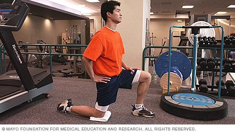
Brace your core and bend your knees to lower down into a split squat. Your left knee should ideally form a 90-degree angle so that your thigh is parallel to the ground, and your right knee is hovering above the floor. (Quick position check: your left foot should be stepped out far enough that you can do this without letting your left knee go past your left toes—if you can't, hop your left foot out a bit farther away from the bench.)
^ Jump up to: a b c d e f Qaseem, A; Wilt, TJ; McLean, RM; Forciea, MA; Clinical Guidelines Committee of the American College of, Physicians. (4 April 2017). "Noninvasive Treatments for Acute, Subacute, and Chronic Low Back Pain: A Clinical Practice Guideline From the American College of Physicians". Annals of Internal Medicine. 166 (7): 514–530. doi:10.7326/M16-2367. PMID 28192789.
Iliopsoas syndrome, which is also called psoas syndrome or iliopsoas tendonitis, occurs when the iliopsoas muscles are injured. Lower back pain is the most common symptom; however, pain can also occur in the hip, thigh, or leg. The iliopsoas bursa, which is a fluid-filled sac located on the inside of the hip that reduces rubbing and friction, is also likely to become inflamed due to the proximity of the two structures. When this happens, the inflamed bursae will make it difficult to move.

