
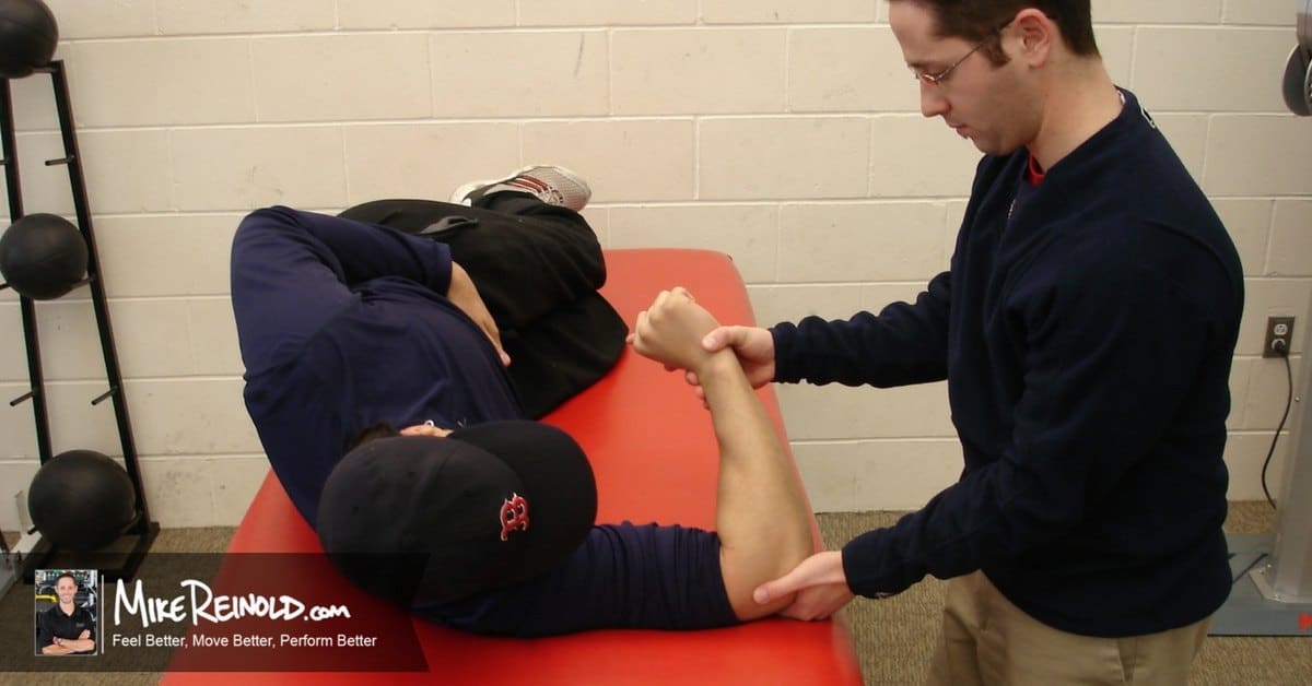
Imaging is indicated when there are red flags, ongoing neurological symptoms that do not resolve, or ongoing or worsening pain.[5] In particular, early use of imaging (either MRI or CT) is recommended for suspected cancer, infection, or cauda equina syndrome.[5] MRI is slightly better than CT for identifying disc disease; the two technologies are equally useful for diagnosing spinal stenosis.[5] Only a few physical diagnostic tests are helpful.[5] The straight leg raise test is almost always positive in those with disc herniation.[5] Lumbar provocative discography may be useful to identify a specific disc causing pain in those with chronic high levels of low back pain.[41] Similarly, therapeutic procedures such as nerve blocks can be used to determine a specific source of pain.[5] Some evidence supports the use of facet joint injections, transforminal epidural injections and sacroilliac injections as diagnostic tests.[5] Most other physical tests, such as evaluating for scoliosis, muscle weakness or wasting, and impaired reflexes, are of little use.[5]

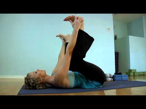
Following any period of prolonged inactivity, a regimen of low-impact exercises is advised. Speed walking, swimming, or stationary bike riding 30 minutes daily can increase muscle strength and flexibility. Yoga also can help stretch and strengthen muscles and improve posture. Consult a physician for a list of low-impact, age-appropriate exercises that are specifically targeted to strengthening lower back and abdominal muscles.
Intradiscal electrothermal therapy (IDET) is a treatment for discs that are cracked or bulging as a result of degenerative disc disease. The procedure involves inserting a catheter through a small incision at the site of the disc in the back. A special wire is passed through the catheter and an electrical current is applied to heat the disc, which helps strengthen the collagen fibers of the disc wall, reducing the bulging and the related irritation of the spinal nerve. IDET is of questionable benefit.
Those are some great stretches! I own a personal training studio in Severna Park, Maryland. Majority of my clients have physical limitations – so it’s important for them to stay flexible. I send these to my clients and even do these exercises for myself. I highly recommend these stretches to anyone, even people without physical limitations. I love the fact these are actually videos and not just stretches because it’s so much easier for people to figure out how to perform the stretches. You guys are the real MVP!

Fitness level: Back pain is more common among people who are not physically fit. Weak back and abdominal muscles may not properly support the spine. “Weekend warriors”—people who go out and exercise a lot after being inactive all week—are more likely to suffer painful back injuries than people who make moderate physical activity a daily habit. Studies show that low-impact aerobic exercise is beneficial for the maintaining the integrity of intervertebral discs.
A traumatic injury, such as from playing sports, car accidents, or a fall can injure tendons, ligaments or muscle resulting in low back pain. Traumatic injury may also cause the spine to become overly compressed, which in turn can cause an intervertebral disc to rupture or herniate, exerting pressure on any of the nerves rooted to the spinal cord. When spinal nerves become compressed and irritated, back pain and sciatica may result.
Apply the above concept to your hips. When you sit, your hips are in a "flexed" position. Therefore, the muscles that flex your hips are in a shortened state. You probably spend at least a third of your day sitting down. Think about how much time those hip flexor muscles stay shortened. A lot. Over time, they become tighter and tighter until you look like the old man in the picture. So unless you want to look like that, perform the stretches shown below.

Epidural steroid injections are a commonly used short-term option for treating low back pain and sciatica associated with inflammation. Pain relief associated with the injections, however, tends to be temporary and the injections are not advised for long-term use. An NIH-funded randomized controlled trial assessing the benefit of epidural steroid injections for the treatment of chronic low back pain associated with spinal stenosis showed that long-term outcomes were worse among those people who received the injections compared with those who did not.
Premkumar et al present evidence that the traditional “red flags” for ominous causes of back pain can be quite misleading. The correlation between red flags and ominous diagnoses is poor, and prone to producing false negatives: that is, no red flags even when there is something more serious than unexplained pain going on. In a survey of almost 10,000 patients “the absence of red flag responses did not meaningfully decrease the likelihood of a red flag diagnosis.“ This is not even remotely a surprise to anyone who paid attention in back pain school, but it’s good to have some harder data on it.
MRI and x-ray for low back pain are surprisingly unreliable,1 because things like bulging discs usually aren’t a deal,2 most back pain goes away on its own,3 and trigger points (“muscle knots”) are common and can be alarmingly intense but aren’t dangerous.4 Most patients are much better off when they feel confident about these things. The power of justified, rational confidence is a huge factor in back pain.5 Sadly, many healthcare professionals continue to perpetuate the idea of fragile backs,6 which undermines that valuable confidence.
Muscle Imbalances – The front of your hips, your hip flexors, are the muscles that will tighten and shorten while you are sitting for hours each day. While you are sitting, the back of your hips, your glutes and your hip extensors, are being overstretched. But just because they are being tightened and stretched respectively, doesn’t benefit either of them. They are also being weakened because of the lack of use of each muscle group.
The iliotibial band is a thickening of the fascia lata, the deep fascia of the thigh. Think of it as a thick long ligament like structure that connects the hip to the lower leg along the outside of the thigh. Tightness in the iliotibial band can cause patellofemoral pain, trochanteric bursitis, and friction syndromes at the knee. This is a hip stretch I commonly prescribe to runners and people suffering from knee pain.
We use cookies and similar technologies to improve your browsing experience, personalize content and offers, show targeted ads, analyze traffic, and better understand you. We may share your information with third-party partners for marketing purposes. To learn more and make choices about data use, visit our Advertising Policy and Privacy Policy. By clicking “Accept and Continue” below, (1) you consent to these activities unless and until you withdraw your consent using our rights request form, and (2) you consent to allow your data to be transferred, processed, and stored in the United States.
When lifting weights, it's important to find out how much weight is appropriate for you. Pariser recommends visiting your physical therapist to discuss how to safely lift weights without injuring your hip. “The lightest weight on the machines might be five or 10 pounds,” Pariser says. “That might be too hard for some people.” A good rule of thumb: Always use a weight that's light enough for you to lift comfortably.
Premkumar et al present evidence that the traditional “red flags” for ominous causes of back pain can be quite misleading. The correlation between red flags and ominous diagnoses is poor, and prone to producing false negatives: that is, no red flags even when there is something more serious than unexplained pain going on. In a survey of almost 10,000 patients “the absence of red flag responses did not meaningfully decrease the likelihood of a red flag diagnosis.“ This is not even remotely a surprise to anyone who paid attention in back pain school, but it’s good to have some harder data on it.
^ Jump up to: a b c d e f g Hughes SP, Freemont AJ, Hukins DW, McGregor AH, Roberts S (October 2012). "The pathogenesis of degeneration of the intervertebral disc and emerging therapies in the management of back pain" (PDF). J Bone Joint Surg Br. 94 (10): 1298–304. doi:10.1302/0301-620X.94B10.28986. PMID 23015552. Archived from the original (PDF) on 4 October 2013. Retrieved 25 June 2013.

The hip joint is where the ball of the thigh bone (femur) joins the pelvis at a socket called the acetabulum. There is cartilage covering both the bone of the femur and the acetabulum of the pelvis in the hip joint. A joint lining tissue, called synovium, surrounds the hip joint. The synovium tissue produces fluid that lubricates the joint and provides nutrients to the cartilage of the joint. The ligaments around the hip joint attach the femur bone to the bony pelvis. There are a number of muscles and tendons that glide around the hip joint. Tiny fluid-filled sacs, called bursae, provide gliding surfaces for muscles and tendons around the hip joint. Major arteries and veins pass the front of the hip joint. The largest nerve of the body, the sciatic nerve, passes behind the hip joint.
Low back pain (LBP) is a common disorder involving the muscles, nerves, and bones of the back.[4] Pain can vary from a dull constant ache to a sudden sharp feeling.[4] Low back pain may be classified by duration as acute (pain lasting less than 6 weeks), sub-chronic (6 to 12 weeks), or chronic (more than 12 weeks).[3] The condition may be further classified by the underlying cause as either mechanical, non-mechanical, or referred pain.[5] The symptoms of low back pain usually improve within a few weeks from the time they start, with 40–90% of people completely better by six weeks.[2]

