Emerging technologies such as X-rays gave physicians new diagnostic tools, revealing the intervertebral disc as a source for back pain in some cases. In 1938, orthopedic surgeon Joseph S. Barr reported on cases of disc-related sciatica improved or cured with back surgery.[100] As a result of this work, in the 1940s, the vertebral disc model of low back pain took over,[99] dominating the literature through the 1980s, aiding further by the rise of new imaging technologies such as CT and MRI.[100] The discussion subsided as research showed disc problems to be a relatively uncommon cause of the pain. Since then, physicians have come to realize that it is unlikely that a specific cause for low back pain can be identified in many cases and question the need to find one at all as most of the time symptoms resolve within 6 to 12 weeks regardless of treatment.[99]
To help you strengthen these important muscles, Miranda put together a list of exercises, below. They include dynamic warm-up moves, meant to activate your hip muscles and prep them for the bigger movements to come; functional moves that train basic movement patterns, like the squat, hip hinge, and lunge; functional plyometric exercises that train explosive power; and a few moves that get you moving in different planes of motion, or directions.
The hip joint is designed to withstand a fair amount of wear and tear, but it’s not indestructible. For example, when you walk, a cushion of cartilage helps prevent friction as the hip bone moves in its socket. With age and use, this cartilage can wear down or become damaged, or the hip bone itself can be fractured during a fall. In fact, more than 300,000 adults over 65 are hospitalized for hip fractures each year, according to the Agency for Healthcare Research and Quality.
For those with pain localized to the lower back due to disc degeneration, fair evidence supports spinal fusion as equal to intensive physical therapy and slightly better than low-intensity nonsurgical measures.[15] Fusion may be considered for those with low back pain from acquired displaced vertebra that does not improve with conservative treatment,[14] although only a few of those who have spinal fusion experience good results.[15] There are a number of different surgical procedures to achieve fusion, with no clear evidence of one being better than the others.[83] Adding spinal implant devices during fusion increases the risks but provides no added improvement in pain or function.[11]
Place a mini band around your ankles and spread your feet about shoulder-width apart. Keeping your legs relatively straight (you want the motion to come from your hips) and toes pointing forward, walk forward 10 steps, then backward 10 steps. Take a short break and then walk to the right 10 steps, then to the left 10 steps. Again, focus on keeping your legs straight and toes pointing forward.
A pinched nerve causes pain, numbness, or tingling in the affected area due to pressure on a nerve. Caral tunnel and sciatica are two examples of conditions caused by a pinched nerve. A pinched nerve is diagnosed by taking a patient history and performing a physical examination. Electromyography may be performed. Treatment for a pinched nerve depends on the underlying cause.
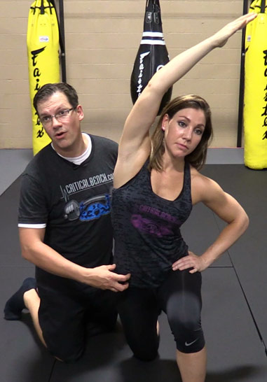
The problem is that these muscles aren't designed to be prime movers—they're designed to support the action of the glutes. Inability of activating the glutes can result in low back pain (low back muscles compensating), hamstring strains (overacting hamstrings), hip pain (resulting from hamstring-dominant hip extension) and knee pain (poor glute medius strength).
Health care professionals diagnose hip pain with a history and physical examination. Physical examination maneuvers, such as internally and externally rotating the hip, can be used to detect pain-aggravating positions. Tenderness can be elicited by palpating over inflamed areas. Straight leg raising can detect signs of sciatica. A health care professional may use imaging studies, including X-rays, CT scans, and MRI scans, to further define the causes of hip pain. Sometimes, nuclear medicine bone scans are used to image inflamed or fractured bone.
To ease the pain and lower your odds of an injury, don’t try to do too much at once. “Start with just 10 minutes,” says Arina Garg, MD, a rheumatology fellow at The Center for Excellence for Arthritis and Rheumatology at the Louisiana University Health Sciences Center. “Every few days, increase that time by 5 to 10 minutes.” Your goal is to work up to 30 minutes of aerobic exercise, 5 days a week.
At the start of the 20th century, physicians thought low back pain was caused by inflammation of or damage to the nerves,[99] with neuralgia and neuritis frequently mentioned by them in the medical literature of the time.[100] The popularity of such proposed causes decreased during the 20th century.[100] In the early 20th century, American neurosurgeon Harvey Williams Cushing increased the acceptance of surgical treatments for low back pain.[14] In the 1920s and 1930s, new theories of the cause arose, with physicians proposing a combination of nervous system and psychological disorders such as nerve weakness (neurasthenia) and female hysteria.[99] Muscular rheumatism (now called fibromyalgia) was also cited with increasing frequency.[100]
^ Jump up to: a b c d e f g Hughes SP, Freemont AJ, Hukins DW, McGregor AH, Roberts S (October 2012). "The pathogenesis of degeneration of the intervertebral disc and emerging therapies in the management of back pain" (PDF). J Bone Joint Surg Br. 94 (10): 1298–304. doi:10.1302/0301-620X.94B10.28986. PMID 23015552. Archived from the original (PDF) on 4 October 2013. Retrieved 25 June 2013.
Luckily, you don’t have to quit your day job or forgo spin class to loosen them up. Simply stretching those hips can get your body back in alignment, increase your mobility (and thus your exercise performance) and maybe even ease pesky back pain, Moore says. “Given the amount of time we sit [each] day and the stress we put our bodies under, hip-opening moves are a necessary party of our daily routine.”
Physical activity can also help you feel better. “Along with boosting your overall health, exercise can improve your OA symptoms” like pain, stiffness, fatigue, and even depression, says Leigh F. Callahan, PhD, associate director of the University of North Carolina Thurston Arthritis Research Center. One study found that people with knee OA who worked out regularly lowered their pain by 12% compared to those who didn’t.
The hip is a common site of osteoarthritis. To help protect the hip joint from "wear and tear," it is important to strengthen the muscles that support it. Your hip also controls the position of your knee, and strengthening your hips may be one component of your rehab program for knee pain. Your physical therapist may also prescribe hip exercises after total hip replacement if you have a hip labrum tear or as part of your hip exercise program for hip pain.
MRI and x-ray for low back pain are surprisingly unreliable,1 because things like bulging discs usually aren’t a deal,2 most back pain goes away on its own,3 and trigger points (“muscle knots”) are common and can be alarmingly intense but aren’t dangerous.4 Most patients are much better off when they feel confident about these things. The power of justified, rational confidence is a huge factor in back pain.5 Sadly, many healthcare professionals continue to perpetuate the idea of fragile backs,6 which undermines that valuable confidence.


3. Tendinitis and bursitis Many tendons around the hip connect the muscles to the joint. These tendons can easily become inflamed if you overuse them or participate in strenuous activities. One of the most common causes of tendinitis at the hip joint, especially in runners, is iliotibial band syndrome — the iliotibial band is the thick span of tissue that runs from the outer rim of your pelvis to the outside of your knee.
THIS TOOL DOES NOT PROVIDE MEDICAL ADVICE. It is intended for general informational purposes only and does not address individual circumstances. It is not a substitute for professional medical advice, diagnosis or treatment and should not be relied on to make decisions about your health. Never ignore professional medical advice in seeking treatment because of something you have read on the WebMD Site. If you think you may have a medical emergency, immediately call your doctor or dial 911.
NINDS-funded studies are contributing to a better understanding of why some people with acute low back pain recover fully while others go on to develop chronic low back pain. Brain imaging studies suggest that people with chronic low back pain have changes in brain structure and function. In one study, people with subacute back pain were followed for one year. Researchers found that certain patterns of functional connectivity across brain networks correlated with the likelihood of pain becoming chronic. The findings suggest that such patterns may help predict who is most likely to transition from subacute to chronic back pain. Other research seeks to determine the role of brain circuits important for emotional and motivational learning and memory in this transition, in order to identify new preventive interventions.
When hip pain comes from muscles, tendons, or ligament injuries, it typically come from overuse syndromes. This can come from overusing the strongest hip muscles in the body such as iliopsoas tendinitis; it can come from tendon and ligament irritations, which typically are involved in snapping hip syndrome. It can come from within the joint, which is more characteristic of hip osteoarthritis. Each of these types of pain present in slightly different ways, which is then the most important part in diagnosing what the cause is by doing a good physical examination.
Antidepressants may be effective for treating chronic pain associated with symptoms of depression, but they have a risk of side effects.[13] Although the antiseizure drugs gabapentin, pregabalin, and topiramate are sometimes used for chronic low back pain evidence does not support a benefit.[79] Systemic oral steroids have not been shown to be useful in low back pain.[1][13] Facet joint injections and steroid injections into the discs have not been found to be effective in those with persistent, non-radiating pain; however, they may be considered for those with persistent sciatic pain.[80] Epidural corticosteroid injections provide a slight and questionable short-term improvement in those with sciatica but are of no long term benefit.[81] There are also concerns of potential side effects.[82]
Using heavy weights: Whether you’re a novice weightlifter or a pro, if you suffer from hip pains you should definitely be putting the kettlebell down! Weight-lifting can place too much stress on your body, especially your knees, lower back and hips. You should be focusing instead on gentle strengthening exercises and stretches in order to support your body as it recovers.
Stand tall with your hips square and bend your right knee, bringing your foot towards your bum. Grab the right foot with your right hand and actively pull the foot closer to your glutes. As you do this, send the right knee down towards the ground and keep both knees together. squeeze your butt to promote a posterior pelvic tilt and hold — then switch sides.