
Deanna is an ACE® certified personal trainer, Balanced Body® Pilates instructor, and NASM® Fitness Nutrition Specialist. She is passionate about inspiring others to lead a healthier lifestyle through fun workouts and healthy food. When she’s not creating new workouts and recipes for her blog The Live Fit Girls she enjoys running with her two dogs and traveling.

There is controversy and scientific uncertainty about trigger points. It’s undeniable that mammals suffer from sensitive spots in our soft tissues … but their nature remains unclear, and the “tiny cramp” theory could be wrong. The tiny cramp theory is formally known as the “expanded integrated hypothesis,” and it has been prominently criticized by Quintner et al (and not many others). However, it’s the mostly widely accepted explanation for now. BACK TO TEXT
The lumbar (or lower back) region is made up of five vertebrae (L1–L5), sometimes including the sacrum. In between these vertebrae are fibrocartilaginous discs, which act as cushions, preventing the vertebrae from rubbing together while at the same time protecting the spinal cord. Nerves come from and go to the spinal cord through specific openings between the vertebrae, providing the skin with sensations and messages to muscles. Stability of the spine is provided by the ligaments and muscles of the back and abdomen. Small joints called facet joints limit and direct the motion of the spine.[30]
Avascular necrosis (also called osteonecrosis). This condition happens when blood flow to the hip bone slows and the bone tissue dies. Although it can affect other bones, avascular necrosis most often happens in the hip. It can be caused by a hip fracture or dislocation, or from the long-term use of high-dose steroids (such as prednisone), among other causes.

Stop listening to other people’s horror stories. You know the scenario: You are bent over in obvious pain, waiting to see the doctor, and the person next to you tells you a 10-minute tale of how their Uncle Gordon had low back pain that required injections and surgery. But the pain still didn’t go away. Stop listening to these terrible stories. Most low back pain is short-lived and can be managed quite effectively with exercise and postural correction. Of course, some low back conditions are serious and require surgery, but that is a conversation you should have with your doctor, not the guy in the waiting room.
If you’re someone who’s got a good stretching routine down, both before and after a HIIT workout, bike ride or run, odds are the discomfort you feel indicates that there are grounds for more strengthening exercises. It’s important to add that many yogis with extremely flexible hips run into overstretching injuries like hip flexor strains. But, these injuries aren’t just limited to the uber-flexible. Runners, cyclists, and Stairmaster lovers might strain these muscles due to frequent overuse. We’ll share a hip flexor workout below but, first up, some stretches.
Spondylolisthesis. This condition occurs when one vertebra slips over the adjacent one. There are 5 types of spondylolisthesis but the most common are secondary to a defect or fracture of the pars (between the facet joints) or mechanical instability of the facet joints (degenerative). The pain can be caused by instability (back) or compression of the nerves (leg).

Lumbar radiculopathy: Lumbar radiculopathy is nerve irritation that is caused by damage to the discs between the vertebrae. Damage to the disc occurs because of degeneration ("wear and tear") of the outer ring of the disc, traumatic injury, or both. As a result, the central softer portion of the disc can rupture (herniate) through the outer ring of the disc and abut the spinal cord or its nerves as they exit the bony spinal column. This rupture is what causes the commonly recognized "sciatica" pain of a herniated disc that shoots from the low back and buttock down the leg. Sciatica can be preceded by a history of localized low-back aching or it can follow a "popping" sensation and be accompanied by numbness and tingling. The pain commonly increases with movements at the waist and can increase with coughing or sneezing. In more severe instances, sciatica can be accompanied by incontinence of the bladder and/or bowels. The sciatica of lumbar radiculopathy typically affects only one side of the body, such as the left side or right side, and not both. Lumbar radiculopathy is suspected based on the above symptoms. Increased radiating pain when the lower extremity is lifted supports the diagnosis. Nerve testing (EMG/electromyogramspina bifida 
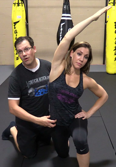
Stand tall with your hips square and bend your right knee, bringing your foot towards your bum. Grab the right foot with your right hand and actively pull the foot closer to your glutes. As you do this, send the right knee down towards the ground and keep both knees together. squeeze your butt to promote a posterior pelvic tilt and hold — then switch sides.
Epidural injections of steroid drugs are frequently used to treat sciatica, despite limited evidence for their effectiveness. Moreover, these treatments are based on the assumption that reducing local inflammation in the vertebral column will relieve pain, but an association between structural abnormalities, inflammation, and sciatica symptoms has not been clearly demonstrated. NINDS-funded researchers are using a new imaging technique that can detect inflammation to better understand what causes chronic sciatica pain and to provide evidence to inform treatment selection.
2016 — More editing, more! Added some better information about pain being a poor indicator, and the role of myofascial trigger points. This article has become extremely busy in the last couple months — about 4,000 readers per day, as described here — so I am really polishing it and making sure that it’s the best possible answer to people’s fears about back pain.

The iliopsoas muscles are a group of two muscles—the psoas muscle and the iliacus muscle—located toward the front of the inner hip. The psoas muscles, in particular, is located in the lumbar (lower) region of the spine and extends through the pelvis to the femur. The iliopsoas muscles are the primary hip flexors, pulling the knee up off the ground when it contracts. Because the psoas muscle is also connected to the spine, it contributes to upright posture, assists in lumbar spine movement, and influences the spine’s curve.
^ Jump up to: a b c d e f Qaseem, A; Wilt, TJ; McLean, RM; Forciea, MA; Clinical Guidelines Committee of the American College of, Physicians. (4 April 2017). "Noninvasive Treatments for Acute, Subacute, and Chronic Low Back Pain: A Clinical Practice Guideline From the American College of Physicians". Annals of Internal Medicine. 166 (7): 514–530. doi:10.7326/M16-2367. PMID 28192789.
I’m not endorsing or saying these stretches are a miracle cure, but I definitely think they’ve helped. I’ve even started to add a few gentle yoga poses and pilates moves which I could so easily do, back in the day, when I was 63 kilos and participated in the greatest oxymoron ever named … a thing called the Fun Run. If I’m not careful, and I keep stretching away each day, I could end up with a 6 pack and back to my 5′ 7″ instead of 5′ 5″.
The lumbar (or lower back) region is made up of five vertebrae (L1–L5), sometimes including the sacrum. In between these vertebrae are fibrocartilaginous discs, which act as cushions, preventing the vertebrae from rubbing together while at the same time protecting the spinal cord. Nerves come from and go to the spinal cord through specific openings between the vertebrae, providing the skin with sensations and messages to muscles. Stability of the spine is provided by the ligaments and muscles of the back and abdomen. Small joints called facet joints limit and direct the motion of the spine.[30]
One of the biggest dangers to your health is constantly sitting for long periods of time which can cause physical and emotional damage. 10 key moves that will help loosen your hip flexor and unlock the power within your body. There is an easy to follow program to unlocking your hip flexors that will strengthen your body, improve your health, and have an all day energy..... https://bit.ly/2HYTPrJ Report
How to: Lie on your back with your right knee bent and foot flat on the floor (a). Extend your left leg up to the ceiling and wrap a strap around the sole of your left foot (b). While holding both ends with your left hand, extend your right arm directly out to the side in order to anchor yourself (c). Slowly let the left leg fall toward the left while keeping your right side grounded. Hold for six to eight breaths, then repeat on the opposite side.
^ Paige, Neil M.; Miake-Lye, Isomi M.; Booth, Marika Suttorp; Beroes, Jessica M.; Mardian, Aram S.; Dougherty, Paul; Branson, Richard; Tang, Baron; Morton, Sally C.; Shekelle, Paul G. (11 April 2017). "Association of Spinal Manipulative Therapy With Clinical Benefit and Harm for Acute Low Back Pain". JAMA. 317 (14): 1451–1460. doi:10.1001/jama.2017.3086. PMC 5470352. PMID 28399251.
Low back pain can be caused by tumors, either benign or malignant, that originate in the bone of the spine or pelvis and spinal cord (primary tumors) and those which originate elsewhere and spread to these areas (metastatic tumors). Symptoms range from localized pain to radiating severe pain and loss of nerve and muscle function (even incontinence of urine and stool) depending on whether or not the tumors affect the nervous tissue. Tumors of these areas are detected using imaging tests, such as plain X-rays, nuclear bone scanning, and CAT and MRI scanning.

It is sometimes hard for an aggressive athlete to consider changing training schedules. It is also hard to accept the fact that a serious disease may exist. All athletes who suffer from low back pain should seek medical advice. Some situations might require reducing or stopping athletic activity until the problem is resolved. The body's ability to be active is worth preserving.
Cancel, pause, or adjust your order at any time, hassle free. Your credit card will only be charged when your order ships. The discount applied every time is 15% off. Since it would be weird to subscribe to a kettlebell, the subscriptions and subscription discounts are only for things you'll need often, like supplements, foods, and personal care items. 
As has been highlighted by research presented at the national meeting of the American College of Rheumatology, a very important aspect of the individual evaluation is the patient's own understanding and perception of their particular situation. British researchers found that those who believed that their symptoms had serious consequences on their lives and that they had, or treatments had, little control over their symptoms were more likely to have a poor outcome. This research points out to physicians the importance of addressing the concerns and perceptions that patients have about their condition during the initial evaluations.

Bleeding in the pelvis is rare without significant trauma and is usually seen in patients who are taking blood-thinning medications, such as warfarin (Coumadin). In these patients, a rapid-onset sciatica pain can be a sign of bleeding in the back of the pelvis and abdomen that is compressing the spinal nerves as they exit to the lower extremities. Infection of the pelvis is infrequent but can be a complication of conditions such as diverticulosis, Crohn's disease, ulcerative colitis, pelvic inflammatory disease with infection of the Fallopian tubes or uterus, and even appendicitis. Pelvic infection is a serious complication of these conditions and is often associated with fever, lowering of blood pressure, and a life-threatening state.

Behavioral therapy may be useful for chronic pain.[16] There are several types available, including operant conditioning, which uses reinforcement to reduce undesirable behaviors and increase desirable behaviors; cognitive behavioral therapy, which helps people identify and correct negative thinking and behavior; and respondent conditioning, which can modify an individual's physiological response to pain.[17] The benefit however is small.[91] Medical providers may develop an integrated program of behavioral therapies.[17] The evidence is inconclusive as to whether mindfulness-based stress reduction reduces chronic back pain intensity or associated disability, although it suggests that it may be useful in improving the acceptance of existing pain.[92][93]
Lie on your back with your knees bent and your feet flat on the floor. Tighten your buttocks and lift your hips off the floor. Tighten your abdominal muscles and lift one foot a couple of inches off the floor. Then put it down and lift the other foot a couple of inches, all while remembering to breathe. “It’s like taking alternate steps,” Pariser says. Work up to doing 30 steps at a time.
Electrodiagnostics are procedures that, in the setting of low back pain, are primarily used to confirm whether a person has lumbar radiculopathy. The procedures include electromyography (EMG), nerve conduction studies (NCS), and evoked potential (EP) studies. EMG assesses the electrical activity in a muscle and can detect if muscle weakness results from a problem with the nerves that control the muscles. Very fine needles are inserted in muscles to measure electrical activity transmitted from the brain or spinal cord to a particular area of the body. NCSs are often performed along with EMG to exclude conditions that can mimic radiculopathy. In NCSs, two sets of electrodes are placed on the skin over the muscles. The first set provides a mild shock to stimulate the nerve that runs to a particular muscle. The second set records the nerve’s electrical signals, and from this information nerve damage that slows conduction of the nerve signal can be detected. EP tests also involve two sets of electrodes—one set to stimulate a sensory nerve, and the other placed on the scalp to record the speed of nerve signal transmissions to the brain.
Many athletes-from the weekend warrior to the elite professional athlete-buck up their strength, pop some over-the-counter pain medication, and tolerate the pain for the sake of the game and personal enjoyment. But avoiding medical help can lead to further and more serious injury. Without medical help, the anatomic damage can sometimes lead to permanent exclusion from sporting activities.
For example, your quadriceps muscles are a group of four that are located at the front of the thigh; one of the group members, the rectus femoris flexes the hip, which brings your lower extremity (thigh, lower leg, and foot) forward, in front of you. On the other hand, your hamstring muscles are located at the back of the thigh. When they contract, they extend the lower extremity, bringing it behind you.
The condition is cauda equina syndrome. It involves “acute loss of function of the neurologic elements (nerve roots) of the spinal canal below the termination (conus) of the spinal cord,” where the nerves spread out like a horse (equina) tail. Again, this condition causes symptoms in the “saddle” of the body: butt, groin, inner thighs. BACK TO TEXT
^ Enke, Oliver; New, Heather A.; New, Charles H.; Mathieson, Stephanie; McLachlan, Andrew J.; Latimer, Jane; Maher, Christopher G.; Lin, C.-W. Christine (2 July 2018). "Anticonvulsants in the treatment of low back pain and lumbar radicular pain: a systematic review and meta-analysis". Canadian Medical Association Journal. 190 (26): E786–E793. doi:10.1503/cmaj.171333. PMC 6028270. PMID 29970367.
Gait analysis studies in the elderly show that they typically have a shortened step length. Whether that is a result of tight hip flexors or due to reduced balance, the propensity to walk with shorter steps will itself lead to tightness in hip flexors and anterior joint structures. Hip stretches may be a relatively easy preventative strategy for the elderly with gait abnormalities and may help to prevent falls.
Premkumar et al present evidence that the traditional “red flags” for ominous causes of back pain can be quite misleading. The correlation between red flags and ominous diagnoses is poor, and prone to producing false negatives: that is, no red flags even when there is something more serious than unexplained pain going on. In a survey of almost 10,000 patients “the absence of red flag responses did not meaningfully decrease the likelihood of a red flag diagnosis.“ This is not even remotely a surprise to anyone who paid attention in back pain school, but it’s good to have some harder data on it.
Your hip labrum is a band of cartilage-like tissue that courses around the outer rim of your hip socket. This labrum helps to support the joint and deepen the socket. Sometimes overuse or an injury to your hip can cause a tear in your labrum. A hip labrum tear may result in a condition called femoroacetabular impingement (FAI). When this happens, hip pain may result, and exercises to stretch and stabilize your hip may be performed.
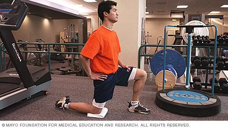
Start in a runner’s lunge with right leg forward, right knee over right ankle and back leg straight. Walk right foot over toward left hand, then drop right shin and thigh to the floor, making sure to keep right knee in line with right hip. Allow left leg to rest on the floor with top of left foot facing down. Take a moment to square your hips to the front of the room. Hold here, or hinge at hips and lower torso toward floor, allowing head to rest on forearms. Hold for at least 30 seconds, then repeat on opposite side. You want to feel a moderate stretch in the outside of the right thigh, but if this pose hurts your knees or feels too uncomfortable, stick with Thread the Needle.
Meanwhile, it’s extremely common for non-life-threatening low back pain to be alarmingly severe and persistent — to have a loud bark! Your doctor may not appreciate how true this is, and may over-react to all persistent low back pain, even without other red flags. In most cases, you shouldn’t let them scare you. Being “freaked out” about persistent back pain is the real threat: it can make low back pain much worse, and much more likely to last even longer (a tragic irony).
Medicine FinderLatest NewsSlip, slap, slop early to save your skinComplementary medicine users have worse cancer survivalVideo: BoredomPut down the tablet; pick up a bookVideo: Lift your mood with some weightsQuicken your step to live longerThis web site is intended for Australian residents and is not a substitute for independent professional advice. Information and interactions contained in this Web site are for information purposes only and are not intended to be used to diagnose, treat, cure or prevent any disease. Further, the accuracy, currency and completeness of the information available on this Web site cannot be guaranteed. Tonic Digital Media Pty Ltd, its affiliates and their respective servants and agents do not accept any liability for any injury, loss or damage incurred by use of or reliance on the information made available via or through myDr whether arising from negligence or otherwise. See Privacy Policy and Disclaimer.2001-2019 myDr.com.au © | All Rights Reserved About UsContact UsDisclaimerPrivacy PolicyAdvertising PolicySitemap

The hip joint is where the ball of the thigh bone (femur) joins the pelvis at a socket called the acetabulum. There is cartilage covering both the bone of the femur and the acetabulum of the pelvis in the hip joint. A joint lining tissue, called synovium, surrounds the hip joint. The synovium tissue produces fluid that lubricates the joint and provides nutrients to the cartilage of the joint. The ligaments around the hip joint attach the femur bone to the bony pelvis. There are a number of muscles and tendons that glide around the hip joint. Tiny fluid-filled sacs, called bursae, provide gliding surfaces for muscles and tendons around the hip joint. Major arteries and veins pass the front of the hip joint. The largest nerve of the body, the sciatic nerve, passes behind the hip joint.
The hip joint is designed to withstand repeated motion and a fair amount of wear and tear. This ball-and-socket joint—the body’s largest ball-and-socket joint—fits together in a way that allows for fluid movement. While many causes of hip pain can arise from the joint itself, there are numerous structures surrounding the hip that can also be the source of pain.
Avascular necrosis (also called osteonecrosis). This condition happens when blood flow to the hip bone slows and the bone tissue dies. Although it can affect other bones, avascular necrosis most often happens in the hip. It can be caused by a hip fracture or dislocation, or from the long-term use of high-dose steroids (such as prednisone), among other causes.
People routinely have no pain despite the presence of obvious arthritic degeneration, herniated discs, and other seemingly serious structural problems like stenosis and spondylolistheses. This surprising contradiction has been made clear by a wide variety of research over the years, but the most notable in recent history is Brinjikji 2015. There are painful spinal problems, of course — which was also shown by Brinjikji et al in a companion paper — but they are mostly more rare and unpredictable than most people suspect, and there are many fascinating examples of people who “should” be in pain but are not, and vice versa. Spinal problems are only one of many ingredients in back pain. BACK TO TEXT
Moist heat may help relax your muscles. Put moist heat on the sore area for 10 to 15 minutes at a time before you do warm-up and stretching exercises. Moist heat includes heat patches or moist heating pads that you can buy at most drugstores, a wet washcloth or towel that has been heated in a microwave or the dryer, or a hot shower. Don’t use heat if you have swelling.
To help you strengthen these important muscles, Miranda put together a list of exercises, below. They include dynamic warm-up moves, meant to activate your hip muscles and prep them for the bigger movements to come; functional moves that train basic movement patterns, like the squat, hip hinge, and lunge; functional plyometric exercises that train explosive power; and a few moves that get you moving in different planes of motion, or directions.
Eleven updates have been logged for this article since publication (2009). All PainScience.com updates are logged to show a long term commitment to quality, accuracy, and currency. more When’s the last time you read a blog post and found a list of many changes made to that page since publication? Like good footnotes, this sets PainScience.com apart from other health websites and blogs. Although footnotes are more useful, the update logs are important. They are “fine print,” but more meaningful than most of the comments that most Internet pages waste pixels on.
Vertebroplasty and kyphoplasty are minimally invasive treatments to repair compression fractures of the vertebrae caused by osteoporosis. Vertebroplasty uses three-dimensional imaging to assist in guiding a fine needle through the skin into the vertebral body, the largest part of the vertebrae. A glue-like bone cement is then injected into the vertebral body space, which quickly hardens to stabilize and strengthen the bone and provide pain relief. In kyphoplasty, prior to injecting the bone cement, a special balloon is inserted and gently inflated to restore height to the vertebral structure and reduce spinal deformity.

