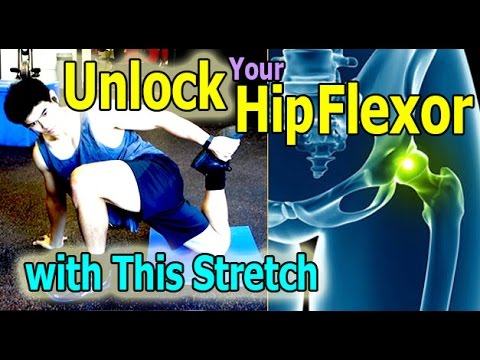
The hip joint is where the ball of the thigh bone (femur) joins the pelvis at a socket called the acetabulum. There is cartilage covering both the bone of the femur and the acetabulum of the pelvis in the hip joint. A joint lining tissue, called synovium, surrounds the hip joint. The synovium tissue produces fluid that lubricates the joint and provides nutrients to the cartilage of the joint. The ligaments around the hip joint attach the femur bone to the bony pelvis. There are a number of muscles and tendons that glide around the hip joint. Tiny fluid-filled sacs, called bursae, provide gliding surfaces for muscles and tendons around the hip joint. Major arteries and veins pass the front of the hip joint. The largest nerve of the body, the sciatic nerve, passes behind the hip joint.
Cycling: The repetitive movements of cycling can place a strain on your hip joints, not to mention that it can also affect your posture! If you really must cycle, make sure your bike is professionally fitted and that you properly warm up and stretch your hip flexors before getting on your bike. I’d recommend choosing an alternative though, or at least speaking to your doctor first!
4. Just swing it. For the front-to-back hip swing stretch, lie on the left side with hips stacked, propped up on the left elbow. Bend the left leg to a 90-degree angle and raise the right leg to hip level with toes pointed. Keep abs tight and swing the right leg all the way in front, then swing it all the way to the back, squeezing the booty along the way. Switch sides.
You may hear a clicking noise when you move your hip, but that sound is not necessarily a hip flexor issue. Siegrist says the clicking isn't generally the hip flexor alone and often comes from a moving part, like the joint. "Maybe there is a loose body in the joint or loose cartilage at the edge of the hip joint that is mechanically getting irritated,” she says.
Sit on floor with knees bent and shins stacked with right leg on top. Use your hand to position right ankle on left knee. Ideally, the right knee will rest on the left thigh, but if your hips are tight, your right knee may point up toward the ceiling (overtime, as your hips become more open, your knee will lower). Keeping your hips squared to the front of the room, hinge at the hips and slowly walk hands slightly forward. If this is enough of a stretch, hold here, or fold your torso over your thighs to go deeper. Hold for at least 30 seconds, then repeat on opposite side.

When we stand, the lower back is functioning to support the weight of the upper body. When we bend, extend, or rotate at the waist, the lower back is involved in the movement. Therefore, injury to the structures important for weight bearing, such as the bony spine, muscles, tendons, and ligaments, often can be detected when the body is standing erect or used in various movements.
But how can you tell? It can be tricky. This is a concise, readable guide to symptoms that need better-safe-than-sorry investigation with your doctor. (It’s basically just a plain English version of clinical guidelines for doctors.9) In other words, this article explains the difference between “dangerous” and “just painful” as clearly as possible. Tables, checklists, and examples ahead.

Doing the bridge exercise in the morning gets your muscles working, activated, and engaged and will help support you the rest of the day, says Humphrey. Lie on your back with your legs bent and your feet flat on the floor, hip-width apart. Press down through your ankles and raise your buttocks off the floor while you tighten your abdominal muscles. Keep your knees aligned with your ankles and aim for a straight line from knees to shoulders, being sure not to arch your back; hold this position for three to five seconds and then slowly lower your buttocks back to the floor. Start with one set of 10 and build up to two or three sets.
Low back pain has been with humans since at least the Bronze Age. The oldest known surgical treatise – the Edwin Smith Papyrus, dating to about 1500 BCE – describes a diagnostic test and treatment for a vertebral sprain. Hippocrates (c. 460 BCE – c. 370 BCE) was the first to use a term for sciatic pain and low back pain; Galen (active mid to late second century CE) described the concept in some detail. Physicians through the end of the first millennium did not attempt back surgery and recommended watchful waiting. Through the Medieval period, folk medicine practitioners provided treatments for back pain based on the belief that it was caused by spirits.[99]

Non-mechanical Disease Processes: Sometimes, non-mechanical disease processes like cancer, kidney stones, or a tumor may cause low back pain. These symptoms are usually, but not always, accompanied by other symptoms like unexpected weight loss, fever, or malaise that indicate a non-mechanical cause of your pain. These diseases are rare, but they can happen, so if your back pain continues for more than a few weeks after physical therapy treatment begins, a visit to your doctor is certainly in order to rule out a sinister problem.
The good news is that there are plenty of good hip stretches out there that you can do to relieve discomfort, decrease tightness, and increase mobility in your hips. Since your hips are involved in so many of the movements you make (both inside and outside of the gym) stretching them is a great way to keep them feeling good and ready to work for you. Add some of the 12 hip stretches Atkins demos below to the end of your workout, or spend 10 minutes each day just doing a few of them, to improve mobility in your hips.
The management of low back pain often includes medications for the duration that they are beneficial. With the first episode of low back pain the hope is a complete cure; however, if the problem becomes chronic, the goals may change to pain management and the recovery of as much function as possible. As pain medications are only somewhat effective, expectations regarding their benefit may differ from reality, and this can lead to decreased satisfaction.[13]
For persistent low back pain, the short-term outcome is also positive, with improvement in the first six weeks but very little improvement after that. At one year, those with chronic low back pain usually continue to have moderate pain and disability.[2] People at higher risk of long-term disability include those with poor coping skills or with fear of activity (2.5 times more likely to have poor outcomes at one year),[96] those with a poor ability to cope with pain, functional impairments, poor general health, or a significant psychiatric or psychological component to the pain (Waddell's signs).[96]
This information is not designed to replace a physician's independent judgment about the appropriateness or risks of a procedure for a given patient. Always consult your doctor about your medical conditions or back problem. SpineUniverse does not provide medical advice, diagnosis or treatment. Use of the SpineUniverse.com site is conditional upon your acceptance of our User Agreement

Emerging technologies such as X-rays gave physicians new diagnostic tools, revealing the intervertebral disc as a source for back pain in some cases. In 1938, orthopedic surgeon Joseph S. Barr reported on cases of disc-related sciatica improved or cured with back surgery.[100] As a result of this work, in the 1940s, the vertebral disc model of low back pain took over,[99] dominating the literature through the 1980s, aiding further by the rise of new imaging technologies such as CT and MRI.[100] The discussion subsided as research showed disc problems to be a relatively uncommon cause of the pain. Since then, physicians have come to realize that it is unlikely that a specific cause for low back pain can be identified in many cases and question the need to find one at all as most of the time symptoms resolve within 6 to 12 weeks regardless of treatment.[99]

Brace your core and bend your knees to lower down into a split squat. Your left knee should ideally form a 90-degree angle so that your thigh is parallel to the ground, and your right knee is hovering above the floor. (Quick position check: your left foot should be stepped out far enough that you can do this without letting your left knee go past your left toes—if you can't, hop your left foot out a bit farther away from the bench.)
Blood tests are not routinely used to diagnose the cause of back pain; however in some cases they may be ordered to look for indications of inflammation, infection, and/or the presence of arthritis. Potential tests include complete blood count, erythrocyte sedimentation rate, and C-reactive protein. Blood tests may also detect HLA-B27, a genetic marker in the blood that is more common in people with ankylosing spondylitis or reactive arthritis (a form of arthritis that occurs following infection in another part of the body, usually the genitourinary tract).
Doing the bridge exercise in the morning gets your muscles working, activated, and engaged and will help support you the rest of the day, says Humphrey. Lie on your back with your legs bent and your feet flat on the floor, hip-width apart. Press down through your ankles and raise your buttocks off the floor while you tighten your abdominal muscles. Keep your knees aligned with your ankles and aim for a straight line from knees to shoulders, being sure not to arch your back; hold this position for three to five seconds and then slowly lower your buttocks back to the floor. Start with one set of 10 and build up to two or three sets.

In the elderly, atherosclerosis can cause weakening of the wall of the large arterial blood vessel (aorta) in the abdomen. This weakening can lead to a bulging (aneurysm) of the aorta wall. While most aneurysms cause no symptoms, some cause a pulsating low back pain. Aneurysms of certain size, especially when enlarging over time, can require surgical repair with a grafting procedure to repair the abnormal portion of the artery.