
Stop focusing on a specific diagnosis. Up to 85% of low back pain can be classified as "non-specific." This means that the origin of your pain cannot be localized to one specific structure or problem. While common diagnostic tests for low back pain can show the bones, discs, and joints with great detail, no test can tell the exact cause of your pain with 100% accuracy.
Nonsteroidal anti-inflammatory drugs (NSAIDS) relieve pain and inflammation and include OTC formulations (ibuprofen, ketoprofen, and naproxen sodium). Several others, including a type of NSAID called COX-2 inhibitors, are available only by prescription. Long-term use of NSAIDs has been associated with stomach irritation, ulcers, heartburn, diarrhea, fluid retention, and in rare cases, kidney dysfunction and cardiovascular disease. The longer a person uses NSAIDs the more likely they are to develop side effects. Many other drugs cannot be taken at the same time a person is treated with NSAIDs because they alter the way the body processes or eliminates other medications.
The hip is a basic ball-and-socket joint. The ball is the femoral head—a knob on the top of the thigh bone—and the socket is an indentation in the pelvic bone. There is cartilage lining the joint (called the labrum) and ligaments that attach the pelvic and thigh bones. Numerous muscles attach around the hip, too, moving the joint through the basic motions of flexion (bending), extension (extending the leg behind you), abduction (lifting the leg away from the body), adduction (moving the leg inward), internal rotation, and external rotation.

Low back pain can cause a wide variety of symptoms and signs depending on the precise cause of the pain as reviewed above. Symptoms that can be associated with low back pain include numbness and/or tingling of the lower extremities, incontinence of urine or stool, inability to walk without worsening pain, lower extremity weakness, atrophy (decreased in size) of the lower extremity muscles, rash, fever, chills, weight loss, abdominal pains, burning on urination, dizziness, joint pain, and fatigue.

Disc degeneration remains a key cause of chronic low back pain and the pain often persists despite surgery. NIH-funded basic science and preclinical studies are investigating molecular-level mechanisms that cause discs in the spine to degenerate, as well as protective mechanisms involved in disc remodeling that may diminish with advancing age. Such studies may help identify future therapeutic strategies to block degenerative mechanisms or promote remodeling processes. NIH also is funding early research on stem cell approaches to promote disc regeneration and rejuvenate cells of the nucleus pulposus, the jelly-like substance in the center of intervertebral discs that loses water content as people age.


The AANS does not endorse any treatments, procedures, products or physicians referenced in these patient fact sheets. This information is provided as an educational service and is not intended to serve as medical advice. Anyone seeking specific neurosurgical advice or assistance should consult his or her neurosurgeon, or locate one in your area through the AANS’ Find a Board-certified Neurosurgeon” online tool. 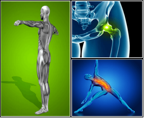
Arthritis. Osteoarthritis and rheumatoid arthritis are among the most common causes of hip pain, especially in older adults. Arthritis leads to inflammation of the hip joint and the breakdown of the cartilage that cushions your hip bones. The pain gradually gets worse. People with arthritis also feel stiffness and have reduced range of motion in the hip.
How to: Lie on your back with your right knee bent and foot flat on the floor (a). Extend your left leg up to the ceiling and wrap a strap around the sole of your left foot (b). While holding both ends with your left hand, extend your right arm directly out to the side in order to anchor yourself (c). Slowly let the left leg fall toward the left while keeping your right side grounded. Hold for six to eight breaths, then repeat on the opposite side.
First and foremost, stop slouching. One of the most common causes of low back pain is poor sitting posture. The strain on the back while sitting in a slouched position can cause excessive pressure on the joints, muscles, and discs, causing pain. Learn to sit with correct posture and maintain that posture at all times to help decrease or eliminate your low back pain. Also be sure your workspace is set up properly at home and at work.
Electrodiagnostics are procedures that, in the setting of low back pain, are primarily used to confirm whether a person has lumbar radiculopathy. The procedures include electromyography (EMG), nerve conduction studies (NCS), and evoked potential (EP) studies. EMG assesses the electrical activity in a muscle and can detect if muscle weakness results from a problem with the nerves that control the muscles. Very fine needles are inserted in muscles to measure electrical activity transmitted from the brain or spinal cord to a particular area of the body. NCSs are often performed along with EMG to exclude conditions that can mimic radiculopathy. In NCSs, two sets of electrodes are placed on the skin over the muscles. The first set provides a mild shock to stimulate the nerve that runs to a particular muscle. The second set records the nerve’s electrical signals, and from this information nerve damage that slows conduction of the nerve signal can be detected. EP tests also involve two sets of electrodes—one set to stimulate a sensory nerve, and the other placed on the scalp to record the speed of nerve signal transmissions to the brain.

Marvelously progressive, concise, and cogent guidelines for physicians on the treatment of low back pain. These guidelines almost entirely “get it right” in my opinion, and are completely consistent with recommendations I’ve been making for years on PainScience.com. They are particularly to be praised for strongly discouraging physicians from ordering imaging tests only “for patients with low back pain when severe or progressive neurologic deficits are present or when serious underlying conditions are suspected.”
Tight hip flexors can also make it harder for your glutes to activate—since they're opposing muscle groups, when one is really tight the other becomes lengthened. When a muscle is more lengthened than it should be, it takes away some of its ability to contract. When your glutes are in this compromised position, it can cause other muscles to do more work than they should, making your workouts less efficient and sometimes, increasing your risk of injury.
Vertebroplasty and kyphoplasty are minimally invasive treatments to repair compression fractures of the vertebrae caused by osteoporosis. Vertebroplasty uses three-dimensional imaging to assist in guiding a fine needle through the skin into the vertebral body, the largest part of the vertebrae. A glue-like bone cement is then injected into the vertebral body space, which quickly hardens to stabilize and strengthen the bone and provide pain relief. In kyphoplasty, prior to injecting the bone cement, a special balloon is inserted and gently inflated to restore height to the vertebral structure and reduce spinal deformity.

