Pain along the inside of the hip may be due to tendinitis or strain of the adductor muscles. Adductors (or inner thigh muscles) pull the leg inward as it is moving forward—the faster the movement, the greater the degree of adduction. Since footprints of a runner are almost single file as opposed to the side-by-side footprints of a walker, there is some degree of adduction occurring during running.
Work on strengthening all of your core muscles and glutes. These muscles work together to give you balance and stability and to help you move through the activities involved in daily living, as well as exercise and sports. When one set of these muscles is weak or tight, it can cause injury or pain in another, so make sure you pay equal attention to all of them.
I’m a runner and started experiencing some familiar tightness in my hip and started getting worse everyday I ran. It’s always gotten sore after running for a long extent for the last 4 years or so. I’m glad I found this page because all of these stretches helped me realize what needed to be stretched and how tight I really was! I hope this will fix my overwhelming soreness. Thank you!

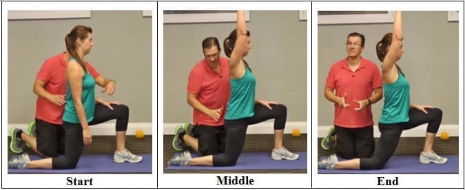
Kneel on your mat with your thighs perpendicular to the floor and tops of your feet facing down. Bring your inner knees together. Slide your feet apart so they are slightly wider than your hips and press the tops of your feet evenly into the mat. Slowly sit down between your feet. Use your hands to turn the top of your thighs inward. Then, lean back onto your forearms and slowly lower torso to floor. Hold for at least 30 seconds.
A pinched nerve causes pain, numbness, or tingling in the affected area due to pressure on a nerve. Caral tunnel and sciatica are two examples of conditions caused by a pinched nerve. A pinched nerve is diagnosed by taking a patient history and performing a physical examination. Electromyography may be performed. Treatment for a pinched nerve depends on the underlying cause.
5. Feel free as a bird. Open up those hips with yoga’s pigeon pose! Start on all fours with hands below the shoulders and knees below the hips. Bring the right knee forward until it touches the right hand and place the leg flat on the ground across the body (the right foot is now on the left side of the body, parallel to the front of the mat). Drop left leg to the ground, and extend it back with toes turned under. Keep the hips level, inhale, and walk hands forward. Exhale, and fold the torso over, lowering elbows to the floor. Stay in this position for 5-10 breaths before coming back up to switch sides.
Even though low back pain can sometimes be treated without major disruption to a person's life, athletes are often reluctant to seek medical help. Many of them deny or minimize complaints in order to avoid consequences, such as: having to decrease activity in order to recover, losing a position or being removed from a team, missing a competition, or letting the team down. Some athletes simply do not want to bother seeing a doctor for pain; they hope it will recover on its own.
A complete medical history and physical exam can usually identify any serious conditions that may be causing the pain. During the exam, a health care provider will ask about the onset, site, and severity of the pain; duration of symptoms and any limitations in movement; and history of previous episodes or any health conditions that might be related to the pain. Along with a thorough back examination, neurologic tests are conducted to determine the cause of pain and appropriate treatment. The cause of chronic lower back pain is often difficult to determine even after a thorough examination.
Model Heather Lin grew up in the deep south but is currently hustling in New York, working at a bank. Whether she is biking home from work, deadlifting, kicking a heavy bag, or pouring all of her effort into a bootcamp class, it's important to her to find time in her busy day to work out. She feels her best when she is strong and energized, and blogs about her health and fitness journey at The Herbivore Warrior.
Congenital bone conditions: Congenital causes (existing from birth) of low back pain include scoliosis and spina bifida. Scoliosis is a sideways (lateral) curvature of the spine that can be caused when one lower extremity is shorter than the other (functional scoliosis) or because of an abnormal architecture of the spine (structural scoliosis). Children who are significantly affected by structural scoliosis may require treatment with bracing and/or surgery to the spine. Adults infrequently are treated surgically but often benefit by support bracing. Spina bifida is a birth defect in the bony vertebral arch over the spinal canal, often with absence of the spinous process. This birth defect most commonly affects the lowest lumbar vertebra and the top of the sacrum. Occasionally, there are abnormal tufts of hair on the skin of the involved area. Spina bifida can be a minor bony abnormality without symptoms. However, the condition can also be accompanied by serious nervous abnormalities of the lower extremities.
The site navigation utilizes arrow, enter, escape, and space bar key commands. Left and right arrows move across top level links and expand / close menus in sub levels. Up and Down arrows will open main level menus and toggle through sub tier links. Enter and space open menus and escape closes them as well. Tab will move on to the next part of the site rather than go through menu items. 
The diagnosis of low back pain involves a review of the history of the illness and underlying medical conditions as well as a physical examination. It is essential that a complete story of the back pain be reviewed including injury history, aggravating and alleviating conditions, associated symptoms (fever, numbness, tingling, incontinence, etc.), as well as the duration and progression of symptoms. Aside from routine abdomen and extremity evaluations, rectal and pelvic examinations may eventually be required as well. Further tests for diagnosis of low back pain can be required including blood and urine tests, plain film X-ray tests, CAT scanning, MRI scanning, bone scanning, and tests of the nerves such as electromyograms (EMG) and nerve conduction velocities (NCV).
This Australian study concluded that “prognosis is moderately optimistic for patients with chronic low back pain,” contradicting the common fear that any low back pain that lasts longer than 6-9 weeks will become a long-term chronic problem. This evidence is the first of its kind, a rarity in low back pain research, a field where almost everything has been studied to death. “Many studies provide good evidence for the prognosis of acute low back pain,” the authors explain. “Relatively few provide good evidence for the prognosis of chronic low back pain.”
While a sharp pain in the hip, groin, pelvis, or thigh is an obvious sign of a hip flexor injury, pain in the lower back and leg are easy to misdiagnose. After all, it’s natural to assume that radiating pain in the leg or lower back originate from these areas. Unfortunately, diagnosing the source of a patient’s pain is not always straightforward. In fact, it’s possible for patients to go years with a misdiagnosed injury.
The hip is a basic ball-and-socket joint. The ball is the femoral head—a knob on the top of the thigh bone—and the socket is an indentation in the pelvic bone. There is cartilage lining the joint (called the labrum) and ligaments that attach the pelvic and thigh bones. Numerous muscles attach around the hip, too, moving the joint through the basic motions of flexion (bending), extension (extending the leg behind you), abduction (lifting the leg away from the body), adduction (moving the leg inward), internal rotation, and external rotation.
^ Jump up to: a b c d e f g Hughes SP, Freemont AJ, Hukins DW, McGregor AH, Roberts S (October 2012). "The pathogenesis of degeneration of the intervertebral disc and emerging therapies in the management of back pain" (PDF). J Bone Joint Surg Br. 94 (10): 1298–304. doi:10.1302/0301-620X.94B10.28986. PMID 23015552. Archived from the original (PDF) on 4 October 2013. Retrieved 25 June 2013.
This Web site provides general educational information on health-related issues and provides access to health-related resources for the convenience of our users. This site and its health-related information and resources are not a substitute for professional medical advice or for the care that patients receive from their physicians or other health care providers.
Bony encroachment: Any condition that results in movement or growth of the vertebrae of the lumbar spine can limit the space (encroachment) for the adjacent spinal cord and nerves. Causes of bony encroachment of the spinal nerves include foraminal narrowing (narrowing of the portal through which the spinal nerve passes from the spinal column, out of the spinal canal to the body, commonly as a result of arthritis), spondylolisthesis (slippage of one vertebra relative to another), and spinal stenosis (compression of the nerve roots or spinal cord by bony spurs or other soft tissues in the spinal canal). Spinal-nerve compression in these conditions can lead to sciatica pain that radiates down the lower extremities. Spinal stenosis can cause lower-extremity pains that worsen with walking and are relieved by resting (mimicking the pains of poor circulation). Treatment of these afflictions varies, depending on their severity, and ranges from rest and exercises to epidural cortisone injections and surgical decompression by removing the bone that is compressing the nervous tissue.
Example: a friend of mine went to the hospital after a motorcycle accident. He’d flown over a car and landed hard on his head. Bizarrely, he was sent home with very little care, and no imaging of his back, even though he was complaining of severe lower back pain. A doctor reassured him that it was just muscle spasms. (This all happened at a hospital that was notorious for being over-crowded and poorly run.) The next day, still in agony, he went to see a doctor at a walk-in clinic, who immediately took him for an x-ray … which identified a serious lumbar fracture and imminent danger of paralysis. He had been lucky to get through the night without disaster! He was placed on a spine board immediately and sent for surgery. The moral of the story? Sometimes, when you’ve had a major trauma and your back really hurts, it’s because your back is broken. BACK TO TEXT
Pain in the hip can result from a number of factors. Sometimes diseases that affect other joints in the body, such as the inflammation resulting from arthritis, can be the cause of pain in the hip. Depending upon the cause of hip pain, the pain may occur when walking, running, or engaging in activity. Trochanteric bursitis is the most common type of hip bursitis and causes pain at the point of the hip.

2016 — More editing, more! Added some better information about pain being a poor indicator, and the role of myofascial trigger points. This article has become extremely busy in the last couple months — about 4,000 readers per day, as described here — so I am really polishing it and making sure that it’s the best possible answer to people’s fears about back pain.

Health Tools Baby Due Date CalculatorBasal Metabolic Rate CalculatorBody Mass Index (BMI) CalculatorCalories Burned CalculatorChild Energy Requirements CalculatorDaily Calcium Requirements CalculatorDaily Fibre Requirements CalculatorIdeal Weight CalculatorInfectious Diseases Exclusion Periods ToolOvulation CalculatorSmoking Cost CalculatorTarget Heart Rate CalculatorWaist-to-hip Ratio Calculator Risk Tests Depression Self-AssessmentErectile Dysfunction ToolMacular Degeneration ToolOsteoporosis Risk TestProstate Symptoms Self-Assessment 
Low back pain can be caused by tumors, either benign or malignant, that originate in the bone of the spine or pelvis and spinal cord (primary tumors) and those which originate elsewhere and spread to these areas (metastatic tumors). Symptoms range from localized pain to radiating severe pain and loss of nerve and muscle function (even incontinence of urine and stool) depending on whether or not the tumors affect the nervous tissue. Tumors of these areas are detected using imaging tests, such as plain X-rays, nuclear bone scanning, and CAT and MRI scanning.
Some of these red flags are much less red than others, especially depending on the circumstances. For instance, “weight loss” is common and often the sign of successful diet! (Well, at least temporarily successful, anyway. 😃) Obviously, if you know of a harmless reason why you have a red flag symptom, it isn’t really a red flag (duh!). But every single actual red flag — in combination with severe low back pain that’s been going on for several weeks — is definitely a good reason to get yourself checked out.
Magnetic resonance imaging (MRI) uses a magnetic force instead of radiation to create a computer-generated image. Unlike x-ray, which shows only bony structures, MRI scans also produce images of soft tissues such as muscles, ligaments, tendons, and blood vessels. An MRI may be ordered if a problem such as infection, tumor, inflammation, disc herniation or rupture, or pressure on a nerve is suspected. MRI is a noninvasive way to identify a condition requiring prompt surgical treatment. However, in most instances, unless there are “red flags” in the history or physical exam, an MRI scan is not necessary during the early phases of low back pain.
Sit on floor with knees bent and shins stacked with right leg on top. Use your hand to position right ankle on left knee. Ideally, the right knee will rest on the left thigh, but if your hips are tight, your right knee may point up toward the ceiling (overtime, as your hips become more open, your knee will lower). Keeping your hips squared to the front of the room, hinge at the hips and slowly walk hands slightly forward. If this is enough of a stretch, hold here, or fold your torso over your thighs to go deeper. Hold for at least 30 seconds, then repeat on opposite side.
Premkumar et al present evidence that the traditional “red flags” for ominous causes of back pain can be quite misleading. The correlation between red flags and ominous diagnoses is poor, and prone to producing false negatives: that is, no red flags even when there is something more serious than unexplained pain going on. In a survey of almost 10,000 patients “the absence of red flag responses did not meaningfully decrease the likelihood of a red flag diagnosis.“ This is not even remotely a surprise to anyone who paid attention in back pain school, but it’s good to have some harder data on it.
Back pain can suck the joy out of your days for week, months, even years. It can definitely be “serious” even when it’s not dangerous. I have worked with many truly miserable chronic low back pain patients, and of course the huge economic costs of back pain are cited practically anywhere the subject comes up. But your typical case of chronic low back pain, as nasty as it can be, has never killed anyone.

The hip flexors play an important role in everyday mobility and exercise. Involved in pulling the knee toward the hip, most movements either directly or indirectly use the hip flexors. That’s why even the slightest injury can cause great discomfort. Learn how you can recover from a hip flexor injury, and what precautions can be taken to avoid them.