
Hip flexors. These hardworking muscles are crucial in foundational movements such as sitting, standing, walking and running — they act as a bridge connecting your torso to your lower body. Some muscles in this group can be notoriously weak or tight and those of you who have ever had issues with this part of your body will know the uncomfortable pain of either all too well. There’s a lot of debate in the world of sports science over how much you should strengthen and stretch your hip flexors — we’ll explain.
How to do it: Loop a resistance band around your ankles and lie on your right side, supporting your upper body with your right hand and forearm. Extend both legs out, feet flexed. Brace your abs in tight and lift your top leg up to hip height, rotating your leg to turn your toes down to the floor, keeping tension on the band. Lift your leg slightly higher than hip height, pushing against band, with heel rotated up to the ceiling. Return to hip height. Repeat 20 times quickly and then switch sides.
Coccydynia is an inflammation of the bony area (tailbone or coccyx) located between the buttocks. Coccydynia is associated with pain and tenderness at the tip of the tailbone between the buttocks. Pain is often worsened by sitting. There are many causes of tailbone pain that can mimic coccydynia including: fracture, pilonidal cysts, infection, and sciatica. Treatment methods include medication and rest.
Pain on the outside of the hip is most commonly due to greater trochanteric bursitis. The greater trochanter is the protrusion where the thigh bone juts outward at the base of the neck (which connects the ball to the femur and is the site of hip stress fractures). A lubricating sac (or bursa) lies over the boney protrusion so that the surrounding muscles do not rub directly on the bone. The top region of the iliotibial (IT) band, known as the tensor fascia lata, is commonly involved in greater trochanteric bursitis.
Pain in the hip can result from a number of factors. Sometimes diseases that affect other joints in the body, such as the inflammation resulting from arthritis, can be the cause of pain in the hip. Depending upon the cause of hip pain, the pain may occur when walking, running, or engaging in activity. Trochanteric bursitis is the most common type of hip bursitis and causes pain at the point of the hip.

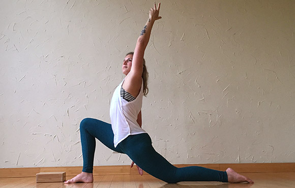
5. Feel free as a bird. Open up those hips with yoga’s pigeon pose! Start on all fours with hands below the shoulders and knees below the hips. Bring the right knee forward until it touches the right hand and place the leg flat on the ground across the body (the right foot is now on the left side of the body, parallel to the front of the mat). Drop left leg to the ground, and extend it back with toes turned under. Keep the hips level, inhale, and walk hands forward. Exhale, and fold the torso over, lowering elbows to the floor. Stay in this position for 5-10 breaths before coming back up to switch sides.
Imagine not being able to climb stairs, bend over, or even walk Changes in hip joint muscle-tendon lengths with mode of locomotion. Riley, P.O., Franz, J., Dicharry, J., et al. Center for Applied Biomechanics, University of Virginia, Charlottesville, VA. Gait & Posture, 2010 Feb; 31 (2): 279-83.. All pretty essential if you ask us! But that’s what our bodies would be like without our hip flexor muscles. Never heard of ‘em? It’s about time we share why they’re so important, how your desk job might be making them weaker (ah!), and the best ways to stretch them out. 
There are cases of low back pain that have alarming causes, but it’s rare. Once in a while back pain is a warning sign of cancer, autoimmune disease, infection, or a handful of other scary culprits.7 Over the age of 55, about one in twenty cases turns out to be a fracture, and one in a hundred is more ominous.8 The further you are from 55, the better your odds.
Paget's disease can be diagnosed on plain X-rays. However, a bone biopsy is occasionally necessary to ensure the accuracy of the diagnosis. Bone scanning is helpful to determine the extent of the disease, which can involve more than one bone area. A blood test, alkaline phosphatase, is useful for diagnosis andmonitoring response to therapy. Treatment options include aspirin, other anti-inflammatory medicines, pain medications, and medications that slow therate of bone turnover, such as calcitonin (Calcimar, Miacalcin), etidronate (Didronel), alendronate (Fosamax), risedronate (Actonel), and pamidronate (Aredia).
Other problems may occur along with low back pain. Chronic low back pain is associated with sleep problems, including a greater amount of time needed to fall asleep, disturbances during sleep, a shorter duration of sleep, and less satisfaction with sleep.[24] In addition, a majority of those with chronic low back pain show symptoms of depression[13] or anxiety.[17]
It is sometimes hard for an aggressive athlete to consider changing training schedules. It is also hard to accept the fact that a serious disease may exist. All athletes who suffer from low back pain should seek medical advice. Some situations might require reducing or stopping athletic activity until the problem is resolved. The body's ability to be active is worth preserving.
This Web site provides general educational information on health-related issues and provides access to health-related resources for the convenience of our users. This site and its health-related information and resources are not a substitute for professional medical advice or for the care that patients receive from their physicians or other health care providers.
^ Enke, Oliver; New, Heather A.; New, Charles H.; Mathieson, Stephanie; McLachlan, Andrew J.; Latimer, Jane; Maher, Christopher G.; Lin, C.-W. Christine (2 July 2018). "Anticonvulsants in the treatment of low back pain and lumbar radicular pain: a systematic review and meta-analysis". Canadian Medical Association Journal. 190 (26): E786–E793. doi:10.1503/cmaj.171333. PMC 6028270. PMID 29970367.
Exercise therapy is effective in decreasing pain and improving function for those with chronic low back pain.[50] It also appears to reduce recurrence rates for as long as six months after the completion of program[61] and improves long-term function.[57] There is no evidence that one particular type of exercise therapy is more effective than another.[62] The Alexander technique appears useful for chronic back pain,[63] and there is tentative evidence to support the use of yoga.[64] Transcutaneous electrical nerve stimulation (TENS) has not been found to be effective in chronic low back pain.[65] Evidence for the use of shoe insoles as a treatment is inconclusive.[51] Peripheral nerve stimulation, a minimally-invasive procedure, may be useful in cases of chronic low back pain that do not respond to other measures, although the evidence supporting it is not conclusive, and it is not effective for pain that radiates into the leg.[66]

Physician specialties that evaluate and treat low back pain range from generalists to subspecialists.These specialties include emergency medicine physicians, general medicine, family medicine, internal medicine, gynecology, spine surgeons (orthopaedics and neurosurgery), rheumatology, pain management, and physiatry. Other health care providers for low back pain include physical therapists, chiropractors, massage therapists, psychologists, and acupuncturists.
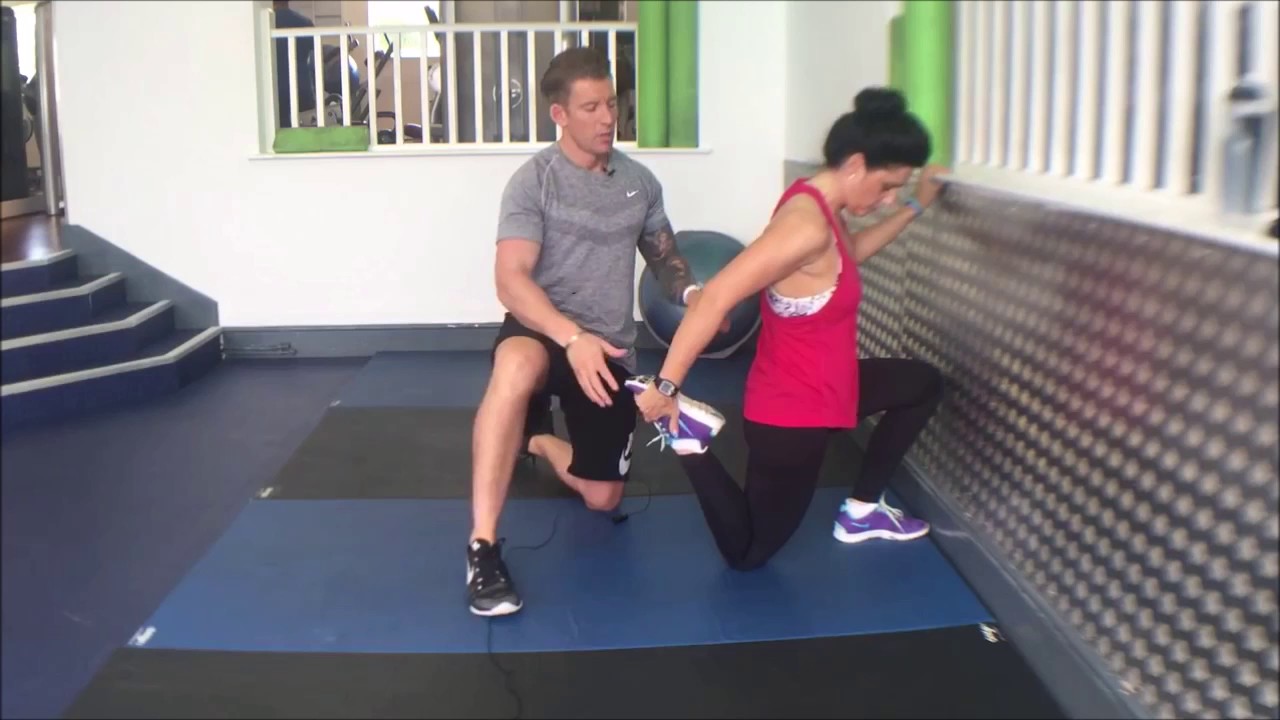
Those are some great stretches! I own a personal training studio in Severna Park, Maryland. Majority of my clients have physical limitations – so it’s important for them to stay flexible. I send these to my clients and even do these exercises for myself. I highly recommend these stretches to anyone, even people without physical limitations. I love the fact these are actually videos and not just stretches because it’s so much easier for people to figure out how to perform the stretches. You guys are the real MVP! 
The information provided herein should not be used during any medical emergency or for the diagnosis or treatment of any medical condition. A licensed physician should be consulted for diagnosis and treatment of any and all medical conditions. Call 911 for all medical emergencies. Links to other sites are provided for information only -- they do not constitute endorsements of those other sites. Copyright 1997-2019, A.D.A.M., Inc. Duplication for commercial use must be authorized in writing by ADAM Health Solutions.
AAOS does not endorse any treatments, procedures, products, or physicians referenced herein. This information is provided as an educational service and is not intended to serve as medical advice. Anyone seeking specific orthopaedic advice or assistance should consult his or her orthopaedic surgeon, or locate one in your area through the AAOS Find an Orthopaedist program on this website.
How to: Lie on your back with your right knee bent and foot flat on the floor (a). With your left leg fully extended, press into your right foot to shift onto your left hip. This is your starting position (b). Then, squeeze your right glutes to press your left hip open until you feel a stretch, pause, then return to start. That’s one rep (c). Perform six to eight reps, then repeat on the opposite side.
Contact sports: It should go without saying; all contact sports should be firmly off your to-do list! That also includes sports than involve contact with another object like tennis or gold! Not only do these forms of exercise make you vulnerable to further injury, the rigorous movements required can place your hip joint under too much stress. Try and give the footie a miss for now and instead focus on other exercises you can do with your friends such as swimming or yoga!
The AANS does not endorse any treatments, procedures, products or physicians referenced in these patient fact sheets. This information is provided as an educational service and is not intended to serve as medical advice. Anyone seeking specific neurosurgical advice or assistance should consult his or her neurosurgeon, or locate one in your area through the AANS’ Find a Board-certified Neurosurgeon” online tool.
If surgery is recommended, neurosurgeons have a variety of options available to help relieve pressure on the nerve roots. If there are several nerve roots and discs causing the pain or if there is degeneration and instability in the spinal column, the neurosurgeon may opt to fuse the vertebrae together with bone grafts and stabilize the vertebrae with instrumentation, including metal plates, screws, rods and cages. A successful fusion will prevent the disc from bulging or herniating again. Following a fusion procedure, a patient may gain restored mobility in the back, including the ability to bend over. He or she will most likely experience more mobility after surgery than before. In addition, the patient may require postoperative physical therapy.
Model Heather Lin grew up in the deep south but is currently hustling in New York, working at a bank. Whether she is biking home from work, deadlifting, kicking a heavy bag, or pouring all of her effort into a bootcamp class, it's important to her to find time in her busy day to work out. She feels her best when she is strong and energized, and blogs about her health and fitness journey at The Herbivore Warrior.
If surgery is recommended, neurosurgeons have a variety of options available to help relieve pressure on the nerve roots. If there are several nerve roots and discs causing the pain or if there is degeneration and instability in the spinal column, the neurosurgeon may opt to fuse the vertebrae together with bone grafts and stabilize the vertebrae with instrumentation, including metal plates, screws, rods and cages. A successful fusion will prevent the disc from bulging or herniating again. Following a fusion procedure, a patient may gain restored mobility in the back, including the ability to bend over. He or she will most likely experience more mobility after surgery than before. In addition, the patient may require postoperative physical therapy.
Discography may be used when other diagnostic procedures fail to identify the cause of pain. This procedure involves the injection of a contrast dye into a spinal disc thought to be causing low back pain. The fluid’s pressure in the disc will reproduce the person’s symptoms if the disc is the cause. The dye helps to show the damaged areas on CT scans taken following the injection. Discography may provide useful information in cases where people are considering lumbar surgery or when their pain has not responded to conventional treatments.

