
NINDS health-related material is provided for information purposes only and does not necessarily represent endorsement by or an official position of the National Institute of Neurological Disorders and Stroke or any other Federal agency. Advice on the treatment or care of an individual patient should be obtained through consultation with a physician who has examined that patient or is familiar with that patient's medical history.
Workers who experience acute low back pain as a result of a work injury may be asked by their employers to have x-rays.[102] As in other cases, testing is not indicated unless red flags are present.[102] An employer's concern about legal liability is not a medical indication and should not be used to justify medical testing when it is not indicated.[102] There should be no legal reason for encouraging people to have tests which a health care provider determines are not indicated.[102]
Congenital bone conditions: Congenital causes (existing from birth) of low back pain include scoliosis and spina bifida. Scoliosis is a sideways (lateral) curvature of the spine that can be caused when one lower extremity is shorter than the other (functional scoliosis) or because of an abnormal architecture of the spine (structural scoliosis). Children who are significantly affected by structural scoliosis may require treatment with bracing and/or surgery to the spine. Adults infrequently are treated surgically but often benefit by support bracing. Spina bifida is a birth defect in the bony vertebral arch over the spinal canal, often with absence of the spinous process. This birth defect most commonly affects the lowest lumbar vertebra and the top of the sacrum. Occasionally, there are abnormal tufts of hair on the skin of the involved area. Spina bifida can be a minor bony abnormality without symptoms. However, the condition can also be accompanied by serious nervous abnormalities of the lower extremities.
The discs are pads that serve as "cushions" between the individual vertebral bodies. They help to minimize the impact of stress forces on the spinal column. Each disc is designed like a jelly donut with a central, softer component (nucleus pulposus) and a surrounding, firm outer ring (annulus fibrosus). The central portion of the disc is capable of rupturing (herniating as in a herniated disc) through the outer ring, causing irritation of adjacent nervous tissue and sciatica as described below. Ligaments are strong fibrous soft tissues that firmly attach bones to bones. Ligaments attach each of the vertebrae to each other and surround each of the discs.

Disc degeneration remains a key cause of chronic low back pain and the pain often persists despite surgery. NIH-funded basic science and preclinical studies are investigating molecular-level mechanisms that cause discs in the spine to degenerate, as well as protective mechanisms involved in disc remodeling that may diminish with advancing age. Such studies may help identify future therapeutic strategies to block degenerative mechanisms or promote remodeling processes. NIH also is funding early research on stem cell approaches to promote disc regeneration and rejuvenate cells of the nucleus pulposus, the jelly-like substance in the center of intervertebral discs that loses water content as people age.

So if you’re doing abs exercises and you feel like your hip flexors are putting in more work, refocus and dial in on the muscles you’re trying to target. It might sound trite to just “think” about a muscle working as you’re doing an exercise (for example, thinking about your abs contracting as you do a sit-up), but it might actually prevent you from mindlessly grinding out reps with poor form.
If you’re worried you’re headed toward a surgeon’s office, there might be hope. According to the Arthritis Foundation, the best way to avoid hip replacement surgery is to get active in an exercise program. In a study, people who participated in an exercise program for 12 weeks were 44 percent less likely to need joint-replacement surgery six years later than those who did not exercise.
"Lower back pain is the most common musculoskeletal ailment in the U.S., and can often be mitigated by strengthening the core musculature," Blake Dircksen, D.P.T., C.S.C.S., a physical therapist at Bespoke Treatments New York, tells SELF. "The 'core' is a cylinder of abdominal and back muscles that wraps around the body like a corset," Dircksen explains. (The glutes are also considered a part of the core, since they connect to the pelvis and ultimately the back and abdominal muscles.) As with any muscles, by strengthening them, you will increase the amount of weight your lower back can comfortably move, which means it will be better equipped to handle the same stress from your workouts and everyday life without getting as achey.
I think you should mention that for some people, stretching is not the solution and that it will deteriorate their posture. Some people need stretching, but most people I know need to strengthen their "overstretched" hip flexors. Many people can't do a single hanging leg raise. Check this site if you want to know more about the importance of hip flexors bit.ly/Unlock_Your_Hip_Flexor Report

5. Feel free as a bird. Open up those hips with yoga’s pigeon pose! Start on all fours with hands below the shoulders and knees below the hips. Bring the right knee forward until it touches the right hand and place the leg flat on the ground across the body (the right foot is now on the left side of the body, parallel to the front of the mat). Drop left leg to the ground, and extend it back with toes turned under. Keep the hips level, inhale, and walk hands forward. Exhale, and fold the torso over, lowering elbows to the floor. Stay in this position for 5-10 breaths before coming back up to switch sides.
Radiofrequency denervation is a procedure using electrical impulses to interrupt nerve conduction (including the conduction of pain signals). Using x-ray guidance, a needle is inserted into a target area of nerves and a local anesthetic is introduced as a way of confirming the involvement of the nerves in the person’s back pain. Next, the region is heated, resulting in localized destruction of the target nerves. Pain relief associated with the technique is temporary and the evidence supporting this technique is limited.
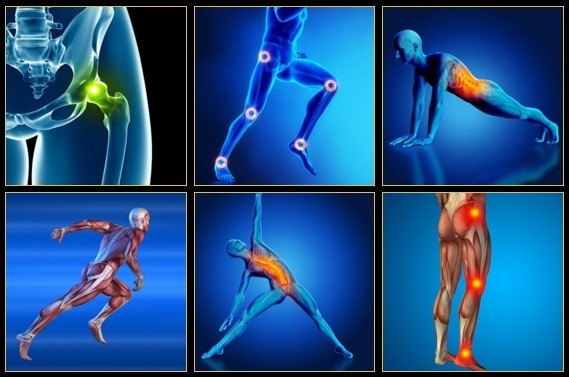
Pain on the outside of the hip is most commonly due to greater trochanteric bursitis. The greater trochanter is the protrusion where the thigh bone juts outward at the base of the neck (which connects the ball to the femur and is the site of hip stress fractures). A lubricating sac (or bursa) lies over the boney protrusion so that the surrounding muscles do not rub directly on the bone. The top region of the iliotibial (IT) band, known as the tensor fascia lata, is commonly involved in greater trochanteric bursitis.
The materials and information provided in this presentation, document and/or any other communication (“Communication”) from Onnit Labs, Inc. or any related entity or person (collectively “Onnit”) are strictly for informational purposes only and are not intended for use as diagnosis, prevention or treatment of a health problem or as a substitute for consulting a qualified medical professional. Some of the concepts presented herein may be theoretical.
Vertebroplasty and kyphoplasty are minimally invasive treatments to repair compression fractures of the vertebrae caused by osteoporosis. Vertebroplasty uses three-dimensional imaging to assist in guiding a fine needle through the skin into the vertebral body, the largest part of the vertebrae. A glue-like bone cement is then injected into the vertebral body space, which quickly hardens to stabilize and strengthen the bone and provide pain relief. In kyphoplasty, prior to injecting the bone cement, a special balloon is inserted and gently inflated to restore height to the vertebral structure and reduce spinal deformity.
At the start of the 20th century, physicians thought low back pain was caused by inflammation of or damage to the nerves,[99] with neuralgia and neuritis frequently mentioned by them in the medical literature of the time.[100] The popularity of such proposed causes decreased during the 20th century.[100] In the early 20th century, American neurosurgeon Harvey Williams Cushing increased the acceptance of surgical treatments for low back pain.[14] In the 1920s and 1930s, new theories of the cause arose, with physicians proposing a combination of nervous system and psychological disorders such as nerve weakness (neurasthenia) and female hysteria.[99] Muscular rheumatism (now called fibromyalgia) was also cited with increasing frequency.[100]
The treatment of lumbar strain consists of resting the back (to avoid reinjury), medications to relieve pain and muscle spasm, local heat applications, massage, and eventual (after the acute episode resolves) reconditioning exercises to strengthen the low back and abdominal muscles. Initial treatment at home might include heat application, acetaminophen (Tylenol) or ibuprofen (Advil, Motrin), and avoiding reinjury and heavy lifting. Prescription medications that are sometimes used for acute low back pain include anti-inflammatory medications, such as sulindac (Clinoril), naproxen (Naprosyn), and ketorolac (Toradol) by injection or by mouth, muscle relaxants, such as carisoprodol (Soma), cyclobenzaprine (Flexeril), methocarbamol (Robaxin), and metaxalone (Skelaxin), as well as analgesics, such as tramadol (Ultram).
Kidneys — The kidneys are a matched pair. One painful kidney can cause back pain on one side or the other. Kidney pain can feel like back pain, and may occur on only one side. It is usually quite lateral, and just barely low enough to qualify as “low” back pain. However, when kidney stones descend through the ureters, they can cause (terrible) pain in the low back. Kidney stone pain is often so severe and develops so rapidly that it isn’t mistaken for a back pain problem.
Spinal fusion eliminates motion between vertebral segments. It is an option when motion is the source of pain. For example, your doctor may recommend spinal fusion if you have spinal instability, a curvature (scoliosis), or severe degeneration of one or more of your disks. The theory is that if the painful spine segments do not move, they should not hurt.
Pregnancy commonly leads to low back pain by mechanically stressing the lumbar spine (changing the normal lumbar curvature) and by the positioning of the baby inside of the abdomen. Additionally, the effects of the female hormone estrogen and the ligament-loosening hormone relaxin may contribute to loosening of the ligaments and structures of the back. Pelvic-tilt exercises and stretches are often recommended for relieving this pain. Women are also recommended to maintain physical conditioning during pregnancy according to their doctors' advice. Natural labor can also cause low back pain.
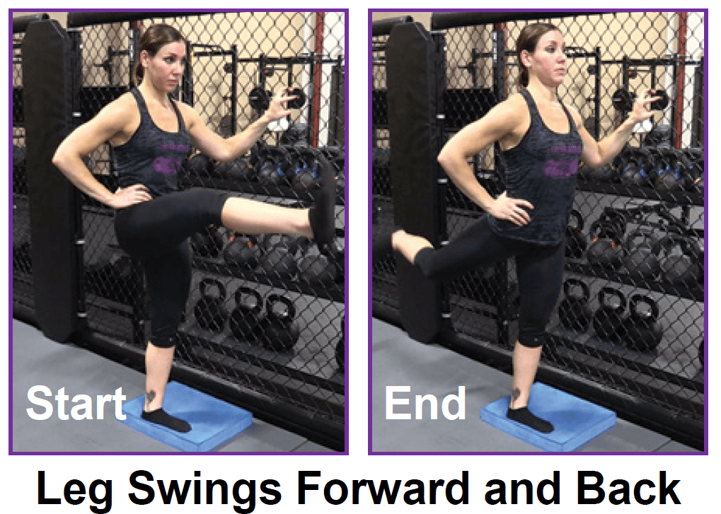
Place a mini band around your ankles and spread your feet about shoulder-width apart. Keeping your legs relatively straight (you want the motion to come from your hips) and toes pointing forward, walk forward 10 steps, then backward 10 steps. Take a short break and then walk to the right 10 steps, then to the left 10 steps. Again, focus on keeping your legs straight and toes pointing forward.

Start kneeling on your mat with knees hip-width apart and hips directly over knees. Press your shins and the tops of your feet into the mat. Bring your hands to your low back, fingers pointing down, and rest palms above glutes. Inhale and lift your chest, and then slowly start to lean your torso back. From here, bring your right hand to rest on your right heel and then your left hand to your left heel. (If you can't reach your heels, turn your toes under; it will be easier to reach your heels in this modification.) Press your thighs forward so they are perpendicular to the floor. Keep your head in a relatively neutral position or, if it doesn't strain your neck, drop it back. Hold for 30 seconds. To come out of the pose, bring your hands to your hips and slowly, leading with your chest, lift your torso as you press the thighs down toward the floor.

