Approximately 15 degrees of hip extension is required to walk normally. If hip flexors are tight then in order to walk, compensatory movement needs to take place through the lower back causing back pain and premature disc degeneration. Like other joints, if we fail to take them through their full range on a regular basis we eventually lose mobility.
The presence of certain signs, termed red flags, indicate the need for further testing to look for more serious underlying problems, which may require immediate or specific treatment.[5][36] The presence of a red flag does not mean that there is a significant problem. It is only suggestive,[37][38] and most people with red flags have no serious underlying problem.[3][1] If no red flags are present, performing diagnostic imaging or laboratory testing in the first four weeks after the start of the symptoms has not been shown to be useful.[5]
Using heavy weights: Whether you’re a novice weightlifter or a pro, if you suffer from hip pains you should definitely be putting the kettlebell down! Weight-lifting can place too much stress on your body, especially your knees, lower back and hips. You should be focusing instead on gentle strengthening exercises and stretches in order to support your body as it recovers.
I’m a runner and started experiencing some familiar tightness in my hip and started getting worse everyday I ran. It’s always gotten sore after running for a long extent for the last 4 years or so. I’m glad I found this page because all of these stretches helped me realize what needed to be stretched and how tight I really was! I hope this will fix my overwhelming soreness. Thank you!
This standard recommendation reinforces the alarming idea that low back pain that lasts longer than a few weeks is Really Bad News. It’s not. It’s a clue. It’s a reason for concern and alertness. But many cases of low back pain that last for 6 weeks will still go away. Once again, see the 2009 research published in the British Medical Journal, which showed that more than 30% of patients with “new” chronic low back pain will still recover without treatment. BACK TO TEXT
Workers who experience acute low back pain as a result of a work injury may be asked by their employers to have x-rays.[102] As in other cases, testing is not indicated unless red flags are present.[102] An employer's concern about legal liability is not a medical indication and should not be used to justify medical testing when it is not indicated.[102] There should be no legal reason for encouraging people to have tests which a health care provider determines are not indicated.[102]
In the vast majority of patients with low back pain, symptoms can be attributed to nonspecific mechanical factors. However, in a much smaller percentage of patients, the cause of back pain may be something more serious, such as cancer, cauda equina syndrome, spinal infection, spinal compression fractures, spinal stress fractures, ankylosing spondylitis, or aneurysm.
Back “spasms” are a largely a myth — there’s no such thing a sustained painful contractions of muscles in otherwise healthy people (see Cramps, Spasms, Tremors & Twitches) — but the kernel of truth in the idea of “spasms” may be the idea of trigger points, which are hypothetical “micro cramps,” tiny patches of painfully contracting muscle. Although this idea is controversial, it is nevertheless one of the most likely explanations for common aches and pains that mostly stick to one area (especially the back) and have no other obvious cause. See Back Pain & Trigger Points. BACK TO TEXT

The hip rotators not only rotate the thigh on the pelvis but more functionally rotate the pelvis on the weight bearing fixed thigh. Activities such as swing a golf club, and even just walking require some rotation of the pelvis on the weight bearing leg. While we don't need that much range of motion to walk, activities such as running, dancing, tennis, and many other sports can require more hip rotation.
With the stakes so high, doesn’t it make sense to do all you can to strengthen and protect your hips? Even if you have arthritis in a hip — the reason for 8 in 10 replacements — you may be able to manage pain with exercise. In a 2017 pilot study published in the Journal of Osteoporosis, women 65 and older who exercised three times a week in a supervised 12-week program reduced arthritic hip pain by over 30 percent, with similar gains in strength, and joint range of motion. The four exercises here will fortify the muscles that surround and support your hips, says trainer Robert Linkul, owner of Be Stronger Fitness in Sacramento, Calif. He advises doing these simple moves, two to three sets of five to 10 reps each, three times a week. Compare how you feel after three weeks.
^ Chou R, Loeser JD, Owens DK, Rosenquist RW, Atlas SJ, Baisden J, Carragee EJ, Grabois M, Murphy DR, Resnick DK, Stanos SP, Shaffer WO, Wall EM, American Pain Society Low Back Pain Guideline Panel (2009). "Interventional therapies, surgery, and interdisciplinary rehabilitation for low back pain: An evidence-based clinical practice guideline from the American Pain Society". Spine. 34 (10): 1066–77. doi:10.1097/BRS.0b013e3181a1390d. PMID 19363457.

The hip joint is designed to withstand a fair amount of wear and tear, but it’s not indestructible. For example, when you walk, a cushion of cartilage helps prevent friction as the hip bone moves in its socket. With age and use, this cartilage can wear down or become damaged, or the hip bone itself can be fractured during a fall. In fact, more than 300,000 adults over 65 are hospitalized for hip fractures each year, according to the Agency for Healthcare Research and Quality.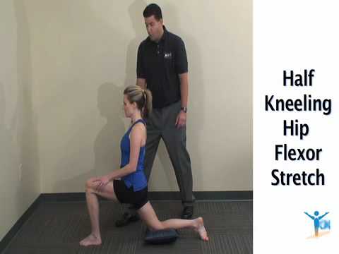
Low back pain that lasts at least one day and limits activity is a common complaint.[7] Globally, about 40% of people have LBP at some point in their lives,[7] with estimates as high as 80% of people in the developed world.[22] Approximately 9 to 12% of people (632 million) have LBP at any given point in time, and nearly one quarter (23.2%) report having it at some point over any one-month period.[7][8] Difficulty most often begins between 20 and 40 years of age.[1] Low back pain is more common among people aged 40–80 years, with the overall number of individuals affected expected to increase as the population ages.[7]
The story of actor Andy Whitfield is a disturbing and educational example of a case that met these conditions — for sure the first two, and probably the third as well if we knew the details. Whitfield was the star of the hit TV show Spartacus (which is worthwhile, but rated very, very R17). The first sign of the cancer that killed him in 2011 was steadily worsening back pain. It’s always hard to diagnose a cancer that starts this way, but Whitfield was in the middle of intense physical training to look the part of history’s most famous gladiator. Back pain didn’t seem unusual at first, and some other symptoms may have been obscured. Weight loss could have even seemed like a training victory at first! It was many long months before he was diagnosed — not until the back pain was severe and constant. A scan finally revealed a large tumour pressing against his spine.
When a muscle contracts, it shortens. Take the biceps for example. Without getting too technical, the biceps are attached at the forearm and shoulder. When your biceps contract, they shorten and bring those two points closer together. When you rest, the muscle returns to its normal length, and the two points move farther away. Constantly contracting your biceps over a long period of time would cause them to get shorter, even at rest.
Hip flexor strains and injuries are often caused by “over doing it” (such as exercising) or periods of prolonged sitting combined with weak hip muscles. While hip flexor strains are typically not serious, they can be quite painful and severely limit your activity and mobility. Airrosti rapidly resolves most hip flexor injuries in as few as 3 visits — without the need for injections, medications, or long periods of rest.

Arthritis: The spondyloarthropathies are inflammatory types of arthritis that can affect the lower back and sacroiliac joints. Examples of spondyloarthropathies include reactive arthritis (Reiter's disease), ankylosing spondylitis, psoriatic arthritis, and the arthritis of inflammatory bowel disease. Each of these diseases can lead to low back pain and stiffness, which is typically worse in the morning. These conditions usually begin in the second and third decades of life. They are treated with medications directed toward decreasing the inflammation. Newer biologic medications have been greatly successful in both quieting the disease and stopping its progression.
Start kneeling on your mat with knees hip-width apart and hips directly over knees. Press your shins and the tops of your feet into the mat. Bring your hands to your low back, fingers pointing down, and rest palms above glutes. Inhale and lift your chest, and then slowly start to lean your torso back. From here, bring your right hand to rest on your right heel and then your left hand to your left heel. (If you can't reach your heels, turn your toes under; it will be easier to reach your heels in this modification.) Press your thighs forward so they are perpendicular to the floor. Keep your head in a relatively neutral position or, if it doesn't strain your neck, drop it back. Hold for 30 seconds. To come out of the pose, bring your hands to your hips and slowly, leading with your chest, lift your torso as you press the thighs down toward the floor.
Hamstring squeeze. Use the machine that works your hamstrings; you will either lie on your stomach or sit with a pad behind your knee. Push against the pad, moving your knee up toward the ceiling or backward (depending on which position you’re in). “In other words, bend your knees,” Pariser says. But to avoid cramps in your hamstring muscles, don’t bend your knee so much that your heels are too close to your buttocks.
As has been highlighted by research presented at the national meeting of the American College of Rheumatology, a very important aspect of the individual evaluation is the patient's own understanding and perception of their particular situation. British researchers found that those who believed that their symptoms had serious consequences on their lives and that they had, or treatments had, little control over their symptoms were more likely to have a poor outcome. This research points out to physicians the importance of addressing the concerns and perceptions that patients have about their condition during the initial evaluations.

I’m not endorsing or saying these stretches are a miracle cure, but I definitely think they’ve helped. I’ve even started to add a few gentle yoga poses and pilates moves which I could so easily do, back in the day, when I was 63 kilos and participated in the greatest oxymoron ever named … a thing called the Fun Run. If I’m not careful, and I keep stretching away each day, I could end up with a 6 pack and back to my 5′ 7″ instead of 5′ 5″.

So, who cares right? Wrong. Everyone has seen that little old man walking with a cane, hunched over almost to the point of staring at the ground. Do you think he always walked like that? I'd bet you he didn't. Maybe he had an injury that never healed properly, or maybe after spending years and years in a similar position, his body became tighter and tighter until eventually he ended up bent over.
Bony encroachment: Any condition that results in movement or growth of the vertebrae of the lumbar spine can limit the space (encroachment) for the adjacent spinal cord and nerves. Causes of bony encroachment of the spinal nerves include foraminal narrowing (narrowing of the portal through which the spinal nerve passes from the spinal column, out of the spinal canal to the body, commonly as a result of arthritis), spondylolisthesis (slippage of one vertebra relative to another), and spinal stenosis (compression of the nerve roots or spinal cord by bony spurs or other soft tissues in the spinal canal). Spinal-nerve compression in these conditions can lead to sciatica pain that radiates down the lower extremities. Spinal stenosis can cause lower-extremity pains that worsen with walking and are relieved by resting (mimicking the pains of poor circulation). Treatment of these afflictions varies, depending on their severity, and ranges from rest and exercises to epidural cortisone injections and surgical decompression by removing the bone that is compressing the nervous tissue.

In diagnosing the causes of hip pain, it’s important to understand hip anatomy. The hip is basically a ball-and-socket joint similar to the shoulder joint and the arms. The hip joint is impressive in that it serves two factors, it is a support structure and also very flexible. Because it is a strong support structure, it tends to get injured. Because it’s very flexible, it similarly can be susceptible to injuries.

So if you’re doing abs exercises and you feel like your hip flexors are putting in more work, refocus and dial in on the muscles you’re trying to target. It might sound trite to just “think” about a muscle working as you’re doing an exercise (for example, thinking about your abs contracting as you do a sit-up), but it might actually prevent you from mindlessly grinding out reps with poor form.
Back pain can suck the joy out of your days for week, months, even years. It can definitely be “serious” even when it’s not dangerous. I have worked with many truly miserable chronic low back pain patients, and of course the huge economic costs of back pain are cited practically anywhere the subject comes up. But your typical case of chronic low back pain, as nasty as it can be, has never killed anyone.
NINDS-funded studies are contributing to a better understanding of why some people with acute low back pain recover fully while others go on to develop chronic low back pain. Brain imaging studies suggest that people with chronic low back pain have changes in brain structure and function. In one study, people with subacute back pain were followed for one year. Researchers found that certain patterns of functional connectivity across brain networks correlated with the likelihood of pain becoming chronic. The findings suggest that such patterns may help predict who is most likely to transition from subacute to chronic back pain. Other research seeks to determine the role of brain circuits important for emotional and motivational learning and memory in this transition, in order to identify new preventive interventions.
In most episodes of low back pain, a specific underlying cause is not identified or even looked for, with the pain believed to be due to mechanical problems such as muscle or joint strain.[1][4] If the pain does not go away with conservative treatment or if it is accompanied by "red flags" such as unexplained weight loss, fever, or significant problems with feeling or movement, further testing may be needed to look for a serious underlying problem.[5] In most cases, imaging tools such as X-ray computed tomography are not useful and carry their own risks.[9][10] Despite this, the use of imaging in low back pain has increased.[11] Some low back pain is caused by damaged intervertebral discs, and the straight leg raise test is useful to identify this cause.[5] In those with chronic pain, the pain processing system may malfunction, causing large amounts of pain in response to non-serious events.[12]
This Australian study concluded that “prognosis is moderately optimistic for patients with chronic low back pain,” contradicting the common fear that any low back pain that lasts longer than 6-9 weeks will become a long-term chronic problem. This evidence is the first of its kind, a rarity in low back pain research, a field where almost everything has been studied to death. “Many studies provide good evidence for the prognosis of acute low back pain,” the authors explain. “Relatively few provide good evidence for the prognosis of chronic low back pain.”
The management of low back pain often includes medications for the duration that they are beneficial. With the first episode of low back pain the hope is a complete cure; however, if the problem becomes chronic, the goals may change to pain management and the recovery of as much function as possible. As pain medications are only somewhat effective, expectations regarding their benefit may differ from reality, and this can lead to decreased satisfaction.[13]
Hip labral tear. This is a rip in the ring of cartilage (called the labrum) that follows the outside rim of the socket of your hip joint. Along with cushioning your hip joint, your labrum acts like a rubber seal or gasket to help hold the ball at the top of your thighbone securely within your hip socket. Athletes and people who perform repetitive twisting movements are at higher risk of developing this problem.
I am a science writer and a former Registered Massage Therapist with a decade of experience treating tough pain cases. I was the Assistant Editor of ScienceBasedMedicine.org for several years. I’ve written hundreds of articles and several books, and I’m known for readable but heavily referenced analysis, with a touch of sass. I am a runner and ultimate player. • more about me • more about PainScience.com
Bleeding in the pelvis is rare without significant trauma and is usually seen in patients who are taking blood-thinning medications, such as warfarin (Coumadin). In these patients, a rapid-onset sciatica pain can be a sign of bleeding in the back of the pelvis and abdomen that is compressing the spinal nerves as they exit to the lower extremities. Infection of the pelvis is infrequent but can be a complication of conditions such as diverticulosis, Crohn's disease, ulcerative colitis, pelvic inflammatory disease with infection of the Fallopian tubes or uterus, and even appendicitis. Pelvic infection is a serious complication of these conditions and is often associated with fever, lowering of blood pressure, and a life-threatening state.
Before recommending exercises, physical therapists evaluate their patients to develop a routine that’s appropriate for their specific condition. Pariser says the following exercises, done at home and at the gym, are generally safe for everyone. “If a patient has already received a total hip replacement, however, certain precautions should be taken,” he says.
Doing the bridge exercise in the morning gets your muscles working, activated, and engaged and will help support you the rest of the day, says Humphrey. Lie on your back with your legs bent and your feet flat on the floor, hip-width apart. Press down through your ankles and raise your buttocks off the floor while you tighten your abdominal muscles. Keep your knees aligned with your ankles and aim for a straight line from knees to shoulders, being sure not to arch your back; hold this position for three to five seconds and then slowly lower your buttocks back to the floor. Start with one set of 10 and build up to two or three sets.
If the pain is still not managed adequately, short term use of opioids such as morphine may be useful.[73][13] These medications carry a risk of addiction, may have negative interactions with other drugs, and have a greater risk of side effects, including dizziness, nausea, and constipation.[13] The effect of long term use of opioids for lower back pain is unknown.[74] Opioid treatment for chronic low back pain increases the risk for lifetime illicit drug use.[75] Specialist groups advise against general long-term use of opioids for chronic low back pain.[13][76] As of 2016, the CDC has released a guideline for prescribed opioid use in the management of chronic pain.[77] It states that opioid use is not the preferred treatment when managing chronic pain due to the excessive risks involved. If prescribed, a person and their clinician should have a realistic plan to discontinue its use in the event that the risks outweigh the benefit.[77]
Place a mini band around your ankles and spread your feet about shoulder-width apart. Keeping your legs relatively straight (you want the motion to come from your hips) and toes pointing forward, walk forward 10 steps, then backward 10 steps. Take a short break and then walk to the right 10 steps, then to the left 10 steps. Again, focus on keeping your legs straight and toes pointing forward.

A pinched nerve causes pain, numbness, or tingling in the affected area due to pressure on a nerve. Caral tunnel and sciatica are two examples of conditions caused by a pinched nerve. A pinched nerve is diagnosed by taking a patient history and performing a physical examination. Electromyography may be performed. Treatment for a pinched nerve depends on the underlying cause.
Eleven updates have been logged for this article since publication (2009). All PainScience.com updates are logged to show a long term commitment to quality, accuracy, and currency. more When’s the last time you read a blog post and found a list of many changes made to that page since publication? Like good footnotes, this sets PainScience.com apart from other health websites and blogs. Although footnotes are more useful, the update logs are important. They are “fine print,” but more meaningful than most of the comments that most Internet pages waste pixels on.

These are really great tips. Just to imform my friends here, my cousin also gave me this link about some other techniches you can use. You have to know exactly what is going on in your body you know. the product is called Panifix, or "Unlock your hip flexor" which Gives You A Practical, Easy-to-follow Program You Can Use To Instantly Release Your Hip Flexors For More Strength, Better Health And All Day Energy. Proven Swipes And Creatives Here:https://tinyurl.com/yd6nbzfh
Following any period of prolonged inactivity, a regimen of low-impact exercises is advised. Speed walking, swimming, or stationary bike riding 30 minutes daily can increase muscle strength and flexibility. Yoga also can help stretch and strengthen muscles and improve posture. Consult a physician for a list of low-impact, age-appropriate exercises that are specifically targeted to strengthening lower back and abdominal muscles.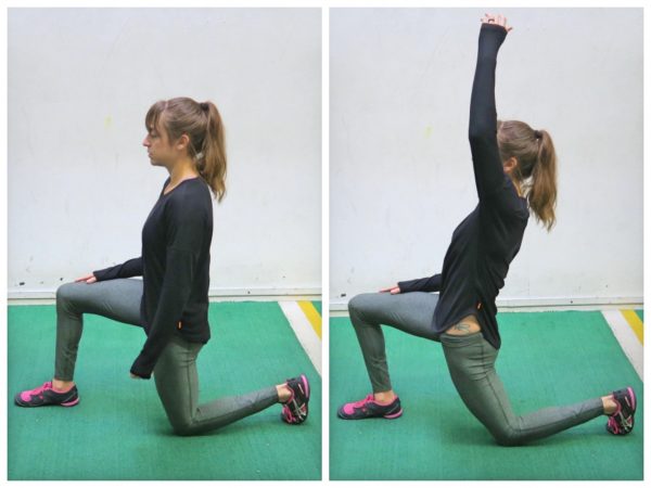
Spinal fusion eliminates motion between vertebral segments. It is an option when motion is the source of pain. For example, your doctor may recommend spinal fusion if you have spinal instability, a curvature (scoliosis), or severe degeneration of one or more of your disks. The theory is that if the painful spine segments do not move, they should not hurt.
The presence of certain signs, termed red flags, indicate the need for further testing to look for more serious underlying problems, which may require immediate or specific treatment.[5][36] The presence of a red flag does not mean that there is a significant problem. It is only suggestive,[37][38] and most people with red flags have no serious underlying problem.[3][1] If no red flags are present, performing diagnostic imaging or laboratory testing in the first four weeks after the start of the symptoms has not been shown to be useful.[5]
Magnetic resonance imaging (MRI) uses a magnetic force instead of radiation to create a computer-generated image. Unlike x-ray, which shows only bony structures, MRI scans also produce images of soft tissues such as muscles, ligaments, tendons, and blood vessels. An MRI may be ordered if a problem such as infection, tumor, inflammation, disc herniation or rupture, or pressure on a nerve is suspected. MRI is a noninvasive way to identify a condition requiring prompt surgical treatment. However, in most instances, unless there are “red flags” in the history or physical exam, an MRI scan is not necessary during the early phases of low back pain.
Congenital bone conditions: Congenital causes (existing from birth) of low back pain include scoliosis and spina bifida. Scoliosis is a sideways (lateral) curvature of the spine that can be caused when one lower extremity is shorter than the other (functional scoliosis) or because of an abnormal architecture of the spine (structural scoliosis). Children who are significantly affected by structural scoliosis may require treatment with bracing and/or surgery to the spine. Adults infrequently are treated surgically but often benefit by support bracing. Spina bifida is a birth defect in the bony vertebral arch over the spinal canal, often with absence of the spinous process. This birth defect most commonly affects the lowest lumbar vertebra and the top of the sacrum. Occasionally, there are abnormal tufts of hair on the skin of the involved area. Spina bifida can be a minor bony abnormality without symptoms. However, the condition can also be accompanied by serious nervous abnormalities of the lower extremities.

^ Jump up to: a b c d e f g Hughes SP, Freemont AJ, Hukins DW, McGregor AH, Roberts S (October 2012). "The pathogenesis of degeneration of the intervertebral disc and emerging therapies in the management of back pain" (PDF). J Bone Joint Surg Br. 94 (10): 1298–304. doi:10.1302/0301-620X.94B10.28986. PMID 23015552. Archived from the original (PDF) on 4 October 2013. Retrieved 25 June 2013.

Electrodiagnostics are procedures that, in the setting of low back pain, are primarily used to confirm whether a person has lumbar radiculopathy. The procedures include electromyography (EMG), nerve conduction studies (NCS), and evoked potential (EP) studies. EMG assesses the electrical activity in a muscle and can detect if muscle weakness results from a problem with the nerves that control the muscles. Very fine needles are inserted in muscles to measure electrical activity transmitted from the brain or spinal cord to a particular area of the body. NCSs are often performed along with EMG to exclude conditions that can mimic radiculopathy. In NCSs, two sets of electrodes are placed on the skin over the muscles. The first set provides a mild shock to stimulate the nerve that runs to a particular muscle. The second set records the nerve’s electrical signals, and from this information nerve damage that slows conduction of the nerve signal can be detected. EP tests also involve two sets of electrodes—one set to stimulate a sensory nerve, and the other placed on the scalp to record the speed of nerve signal transmissions to the brain.

How to do it: Loop a resistance band around your ankles and lie on your right side with your right arm extended on the floor, left hand in front of your body for support. Brace your abs in tight, bring your bottom leg slightly in front of your top leg (let the inside of your top foot rest on the ground), and lift your leg up to the ceiling, keeping your hips stacked. Maintain tension on the band at all times and pulse your bottom leg up and down quickly 20 times. Repeat on the other side.
You may hear a clicking noise when you move your hip, but that sound is not necessarily a hip flexor issue. Siegrist says the clicking isn't generally the hip flexor alone and often comes from a moving part, like the joint. "Maybe there is a loose body in the joint or loose cartilage at the edge of the hip joint that is mechanically getting irritated,” she says.

At the very least, the tension and/or spasm in muscles that cross over the hip and attach onto the pelvis can contribute to imbalance, in terms of how strong and flexible each muscle group is in relation to the others. But muscle imbalance in the hips and the spine may make for pain, limitation and/or posture problems. It can also increase the healing challenge put to you by an existing injury or condition, for example, scoliosis.
Quick anatomy lesson. When we talk about the hips, we're talking about any muscle that crosses over the hip joint, says Laura Miranda D.P.T., M.S.P.T., C.S.C.S., a New York City-based trainer and creator of the Pursuit training program. Which, there are many, including all of the glute muscles, the hamstrings, the inner thigh muscles, and the psoas muscles (deep core muscles that attach your pelvis to your spine). Each of these muscles has some specific roles, but overall, the hip muscles stabilize your pelvis and thighbone as you move. They also allow you to bend at the hips, lift your legs out to the side (abduct), and bring your legs back in toward one another (adduct). Basically, they do a lot, and when they're weak or tight or otherwise not working in an optimal way, you can not only end up with cranky hips, but other body parts may overcompensate and take on too much work—leaving you with other, seemingly unrelated, issues, like knee pain.
Complaints of low back pain are one of the most common reasons people visit doctors.[9][42] For pain that has lasted only a few weeks, the pain is likely to subside on its own.[43] Thus, if a person's medical history and physical examination do not suggest a specific disease as the cause, medical societies advise against imaging tests such as X-rays, CT scans, and MRIs.[42] Individuals may want such tests but, unless red flags are present,[10][44] they are unnecessary health care.[9][43] Routine imaging increases costs, is associated with higher rates of surgery with no overall benefit,[45][46] and the radiation used may be harmful to one's health.[45] Fewer than 1% of imaging tests identify the cause of the problem.[9] Imaging may also detect harmless abnormalities, encouraging people to request further unnecessary testing or to worry.[9] Even so, MRI scans of the lumbar region increased by more than 300% among United States Medicare beneficiaries from 1994 to 2006.[11]
Talmage, J; Belcourt, R; Galper, J; et al. (2011). "Low back disorders". In Kurt T. Hegmann. Occupational medicine practice guidelines : evaluation and management of common health problems and functional recovery in workers (3rd ed.). Elk Grove Village, IL: American College of Occupational and Environmental Medicine. pp. 336, 373, 376–377. ISBN 978-0615452272.

Management of low back pain depends on which of the three general categories is the cause: mechanical problems, non-mechanical problems, or referred pain.[52] For acute pain that is causing only mild to moderate problems, the goals are to restore normal function, return the individual to work, and minimize pain. The condition is normally not serious, resolves without much being done, and recovery is helped by attempting to return to normal activities as soon as possible within the limits of pain.[3] Providing individuals with coping skills through reassurance of these facts is useful in speeding recovery.[1] For those with sub-chronic or chronic low back pain, multidisciplinary treatment programs may help.[53] Initial management with non–medication based treatments is recommended, with NSAIDs used if these are not sufficiently effective.[6]
The good news is that there are plenty of good hip stretches out there that you can do to relieve discomfort, decrease tightness, and increase mobility in your hips. Since your hips are involved in so many of the movements you make (both inside and outside of the gym) stretching them is a great way to keep them feeling good and ready to work for you. Add some of the 12 hip stretches Atkins demos below to the end of your workout, or spend 10 minutes each day just doing a few of them, to improve mobility in your hips.
Simply stand up straight with your feet about shoulder-width apart. Slowly bend your knees and hips, lowering yourself until your knees obscure your toes or you achieve a 90 degree angle. Hold for a count of 5 and then gently resume your original position. This can be a tough one so again, don’t overdo it and hold on to a table if you need a little extra support! Try to repeat between 5-10 times.
Pain in the front of the joint—where the leg attaches to the trunk—is typically caused by hip joint problems. You may also feel pain associated with a hip injury in the lower part of your glutes and the top of the back of your thigh. Other symptoms include the inability to move the leg at the hip, inability to put weight on one leg due to pain at the hip, or swelling around the joint. As runners, you may also experience pain in the hipo joint only when running due to a hip injury.
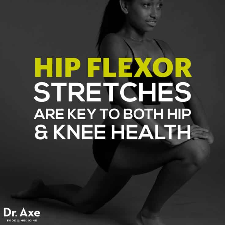
Located deep in the front of the hip and connecting the leg, pelvis, and abdomen, the hip flexors— surprise, surprise— flex the hip. But despite being some of the most powerful muscles in our bodies (with a clearly important role), it’s easy to neglect our poor hip flexors— often without even knowing it. It turns out just working at a desk all day (guilty!) can really weaken hip flexors since they tend to shorten up while in a seated position. This tightness disrupts good posture and is a common cause of lower back pain. Weakened hip flexors can also increase the risk of foot, ankle, and knee injuries (especially among runners) Hip muscle weakness and overuse injuries in recreational runners. Niemuth, P.E., Johnson, R.J., Myers, M.J., et al. Rocky Mountain University of Health Professions, Provo, VT. Clinical Journal of Sport Medicine, 2005 Jan; 15 (1): 14-21.. So be sure to get up, stand up every hour or so! And giving the hip flexors some extra attention is not just about injury prevention. Adding power to workouts, working toward greater flexibility, and getting speedier while running is also, as they say, all in the hips The effect of walking speed on muscle function and mechanical energetics. Neptune, R.R., Sasaki, K., and Kautz, S.A. Department of Mechanical Engineering, The University of Texas, Austin, TX. Gait & Posture, 2008 Jul; 28 (1): 135-43..