Bleeding in the pelvis is rare without significant trauma and is usually seen in patients who are taking blood-thinning medications, such as warfarin (Coumadin). In these patients, a rapid-onset sciatica pain can be a sign of bleeding in the back of the pelvis and abdomen that is compressing the spinal nerves as they exit to the lower extremities. Infection of the pelvis is infrequent but can be a complication of conditions such as diverticulosis, Crohn's disease, ulcerative colitis, pelvic inflammatory disease with infection of the Fallopian tubes or uterus, and even appendicitis. Pelvic infection is a serious complication of these conditions and is often associated with fever, lowering of blood pressure, and a life-threatening state.
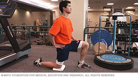
Gait analysis studies in the elderly show that they typically have a shortened step length. Whether that is a result of tight hip flexors or due to reduced balance, the propensity to walk with shorter steps will itself lead to tightness in hip flexors and anterior joint structures. Hip stretches may be a relatively easy preventative strategy for the elderly with gait abnormalities and may help to prevent falls.
Emerging technologies such as X-rays gave physicians new diagnostic tools, revealing the intervertebral disc as a source for back pain in some cases. In 1938, orthopedic surgeon Joseph S. Barr reported on cases of disc-related sciatica improved or cured with back surgery.[100] As a result of this work, in the 1940s, the vertebral disc model of low back pain took over,[99] dominating the literature through the 1980s, aiding further by the rise of new imaging technologies such as CT and MRI.[100] The discussion subsided as research showed disc problems to be a relatively uncommon cause of the pain. Since then, physicians have come to realize that it is unlikely that a specific cause for low back pain can be identified in many cases and question the need to find one at all as most of the time symptoms resolve within 6 to 12 weeks regardless of treatment.[99]

The hip joint is designed to withstand a fair amount of wear and tear, but it’s not indestructible. For example, when you walk, a cushion of cartilage helps prevent friction as the hip bone moves in its socket. With age and use, this cartilage can wear down or become damaged, or the hip bone itself can be fractured during a fall. In fact, more than 300,000 adults over 65 are hospitalized for hip fractures each year, according to the Agency for Healthcare Research and Quality.
Subacute low back pain. Lasting between 6 weeks and 3 months, this type of pain is usually mechanical in nature (such as a muscle strain or joint pain) but is prolonged. At this point, a medical workup may be considered, and is advisable if the pain is severe and limits one’s ability to participate in activities of daily living, sleeping, and working.
There are many causes of hip joint pain. Some hip pain is temporary, while other hip pain can be long-standing or chronic. Causes of hip pain include arthritis, inflammatory and noninflammatory arthritis, fracture, sprain, infectious arthritis (septic arthritis), avascular necrosis, Gaucher's disease, sciatica, muscle strain, iliotibial band syndrome (IT band syndrome), and hematoma.
Deanna is an ACE® certified personal trainer, Balanced Body® Pilates instructor, and NASM® Fitness Nutrition Specialist. She is passionate about inspiring others to lead a healthier lifestyle through fun workouts and healthy food. When she’s not creating new workouts and recipes for her blog The Live Fit Girls she enjoys running with her two dogs and traveling.
The multifidus muscles run up and down along the back of the spine, and are important for keeping the spine straight and stable during many common movements such as sitting, walking and lifting.[12] A problem with these muscles is often found in someone with chronic low back pain, because the back pain causes the person to use the back muscles improperly in trying to avoid the pain.[31] The problem with the multifidus muscles continues even after the pain goes away, and is probably an important reason why the pain comes back.[31] Teaching people with chronic low back pain how to use these muscles is recommended as part of a recovery program.[31]
Electrodiagnostics are procedures that, in the setting of low back pain, are primarily used to confirm whether a person has lumbar radiculopathy. The procedures include electromyography (EMG), nerve conduction studies (NCS), and evoked potential (EP) studies. EMG assesses the electrical activity in a muscle and can detect if muscle weakness results from a problem with the nerves that control the muscles. Very fine needles are inserted in muscles to measure electrical activity transmitted from the brain or spinal cord to a particular area of the body. NCSs are often performed along with EMG to exclude conditions that can mimic radiculopathy. In NCSs, two sets of electrodes are placed on the skin over the muscles. The first set provides a mild shock to stimulate the nerve that runs to a particular muscle. The second set records the nerve’s electrical signals, and from this information nerve damage that slows conduction of the nerve signal can be detected. EP tests also involve two sets of electrodes—one set to stimulate a sensory nerve, and the other placed on the scalp to record the speed of nerve signal transmissions to the brain.
If you are experiencing true numbness14 around the groin and buttocks and/or failure of bladder or bowel control, please consider it a serious emergency — do not wait to see if it goes away. These symptoms indicate spinal cord injury or compression15 and require immediate medical attention. (Few people will have symptoms like this without having already decided it’s an emergency, but I have to cover all the bases here.)
If you have arthritis or bursitis, you’ve probably noticed that exercise can actually help relieve your pain. But when is hip pain a sign that you should stop exercising or doing a certain activity? If your hip starts to hurt during a particular exercise and lingers for hours or days afterward, that’s a sign that your joint needs to rest, notes Humphrey. It’s normal to feel some soreness the day after exercising, but the pain shouldn’t persist or become worse. Also, if you experience a sharp or shooting pain, stop the activity immediately and talk to your doctor or physical therapist.
The multifidus muscles run up and down along the back of the spine, and are important for keeping the spine straight and stable during many common movements such as sitting, walking and lifting.[12] A problem with these muscles is often found in someone with chronic low back pain, because the back pain causes the person to use the back muscles improperly in trying to avoid the pain.[31] The problem with the multifidus muscles continues even after the pain goes away, and is probably an important reason why the pain comes back.[31] Teaching people with chronic low back pain how to use these muscles is recommended as part of a recovery program.[31]
I think you should mention that for some people, stretching is not the solution and that it will deteriorate their posture. Some people need stretching, but most people I know need to strengthen their "overstretched" hip flexors. Many people can't do a single hanging leg raise. Check this site if you want to know more about the importance of hip flexors bit.ly/Unlock_Your_Hip_Flexor Report

I am a science writer, former massage therapist, and I was the assistant editor at ScienceBasedMedicine.org for several years. I have had my share of injuries and pain challenges as a runner and ultimate player. My wife and I live in downtown Vancouver, Canada. See my full bio and qualifications, or my blog, Writerly. You might run into me on Facebook or Twitter.

Gait analysis studies in the elderly show that they typically have a shortened step length. Whether that is a result of tight hip flexors or due to reduced balance, the propensity to walk with shorter steps will itself lead to tightness in hip flexors and anterior joint structures. Hip stretches may be a relatively easy preventative strategy for the elderly with gait abnormalities and may help to prevent falls.
There are cases of low back pain that have alarming causes, but it’s rare. Once in a while back pain is a warning sign of cancer, autoimmune disease, infection, or a handful of other scary culprits.7 Over the age of 55, about one in twenty cases turns out to be a fracture, and one in a hundred is more ominous.8 The further you are from 55, the better your odds.
Blood tests are not routinely used to diagnose the cause of back pain; however in some cases they may be ordered to look for indications of inflammation, infection, and/or the presence of arthritis. Potential tests include complete blood count, erythrocyte sedimentation rate, and C-reactive protein. Blood tests may also detect HLA-B27, a genetic marker in the blood that is more common in people with ankylosing spondylitis or reactive arthritis (a form of arthritis that occurs following infection in another part of the body, usually the genitourinary tract).
Premkumar et al present evidence that the traditional “red flags” for ominous causes of back pain can be quite misleading. The correlation between red flags and ominous diagnoses is poor, and prone to producing false negatives: that is, no red flags even when there is something more serious than unexplained pain going on. In a survey of almost 10,000 patients “the absence of red flag responses did not meaningfully decrease the likelihood of a red flag diagnosis.“ This is not even remotely a surprise to anyone who paid attention in back pain school, but it’s good to have some harder data on it.
This game-changing mat is what every yogi wishes they could practice in. Its smooth top layer provides better grip for tricky poses and sweaty hot yoga sessions. The thick fabric supports knees and elbows when you're in plank and pigeon. But what sets this yoga mat apart is its ability to roll up on its own and snap in place. This self-rolling mat also pairs with the Women's Health Amazon Alexa app, which walks you through the flow of the day.
In the elderly, atherosclerosis can cause weakening of the wall of the large arterial blood vessel (aorta) in the abdomen. This weakening can lead to a bulging (aneurysm) of the aorta wall. While most aneurysms cause no symptoms, some cause a pulsating low back pain. Aneurysms of certain size, especially when enlarging over time, can require surgical repair with a grafting procedure to repair the abnormal portion of the artery.