
The AANS does not endorse any treatments, procedures, products or physicians referenced in these patient fact sheets. This information is provided as an educational service and is not intended to serve as medical advice. Anyone seeking specific neurosurgical advice or assistance should consult his or her neurosurgeon, or locate one in your area through the AANS’ Find a Board-certified Neurosurgeon” online tool.
According to a study published in Annals of Internal Medicine, there is strong evidence that yoga can have a short-term effect on treating lower back pain. Yoga involves slow, controlled movements to stretch and strengthen the body. This exercise form also promotes stress relief, which can help reduce tension you may commonly hold in your lower back.
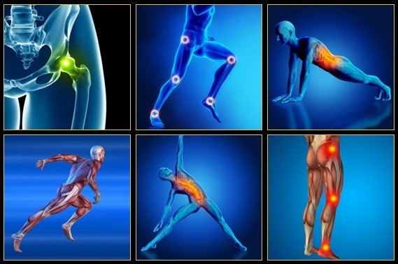
In diagnosing the causes of hip pain, it’s important to understand hip anatomy. The hip is basically a ball-and-socket joint similar to the shoulder joint and the arms. The hip joint is impressive in that it serves two factors, it is a support structure and also very flexible. Because it is a strong support structure, it tends to get injured. Because it’s very flexible, it similarly can be susceptible to injuries.
Their research differs from past studies of chronic low back pain, which tended to focus on patients who already had a well-established track record of long-term problems (in other words, the people who had already drawn the short straw before they were selected for study, and are likely to carry right on feeling rotten). Instead they studied new cases of chronic low back pain, and found that “more than one third” recovered within nine more months. This evidence is a great foundation for more substantive and lasting reassurance for low back pain patients.
Lay on your back on your mat and pull your knees to your chest. Place your hands on the inside arches of your feet and open your knees wider than shoulder-width apart. Keeping your back pressed into the mat as much as possible, press your feet into hands while pulling down on feet, creating resistance. Breathe deeply and hold for at least 30 seconds.

There are a number of ways to classify low back pain with no consensus that any one method is best.[5] There are three general types of low back pain by cause: mechanical back pain (including nonspecific musculoskeletal strains, herniated discs, compressed nerve roots, degenerative discs or joint disease, and broken vertebra), non-mechanical back pain (tumors, inflammatory conditions such as spondyloarthritis, and infections), and referred pain from internal organs (gallbladder disease, kidney stones, kidney infections, and aortic aneurysm, among others).[5] Mechanical or musculoskeletal problems underlie most cases (around 90% or more),[5][34] and of those, most (around 75%) do not have a specific cause identified, but are thought to be due to muscle strain or injury to ligaments.[5][34] Rarely, complaints of low back pain result from systemic or psychological problems, such as fibromyalgia and somatoform disorders.[34]
You could do these moves all together as a single workout, or, as Miranda suggests, split them in half and do the first part one day and the second part another—"but do the warm-up with each one," she says. Those first three moves are meant to not only "wake up" the muscles, but also get your brain ready for the movement patterns to come. For that reason, she says that doing the first three moves "would be a fantastic warm-up before any workout."
When a muscle contracts, it shortens. Take the biceps for example. Without getting too technical, the biceps are attached at the forearm and shoulder. When your biceps contract, they shorten and bring those two points closer together. When you rest, the muscle returns to its normal length, and the two points move farther away. Constantly contracting your biceps over a long period of time would cause them to get shorter, even at rest.
In diagnosing the causes of hip pain, it’s important to understand hip anatomy. The hip is basically a ball-and-socket joint similar to the shoulder joint and the arms. The hip joint is impressive in that it serves two factors, it is a support structure and also very flexible. Because it is a strong support structure, it tends to get injured. Because it’s very flexible, it similarly can be susceptible to injuries.

If low back pain gets worse or does not improve after two to three days of home treatment, contact a primary-care physician. The physician can evaluate the patient and perform a neurological exam in the office to determine which nerve root is being irritated, as well as rule out other serious medical conditions. If there are clear signs that the nerve root is being compressed, a physician can prescribe medications to relieve the pain, swelling and irritation; he or she also may recommend limitation of activities. If these treatment options do not provide relief within two weeks, it may be time to consider other diagnostic studies and possibly surgery.

When intervertebral disks collapse and osteoarthritis develops, your body may respond by growing new bone in your facet joints to help support the vertebrae. Over time, this bone overgrowth (called spurs) can lead to a narrowing of the spinal canal. Osteoarthritis can also cause the ligaments that connect vertebrae to thicken, which can narrow the spinal canal.
Congenital bone conditions: Congenital causes (existing from birth) of low back pain include scoliosis and spina bifida. Scoliosis is a sideways (lateral) curvature of the spine that can be caused when one lower extremity is shorter than the other (functional scoliosis) or because of an abnormal architecture of the spine (structural scoliosis). Children who are significantly affected by structural scoliosis may require treatment with bracing and/or surgery to the spine. Adults infrequently are treated surgically but often benefit by support bracing. Spina bifida is a birth defect in the bony vertebral arch over the spinal canal, often with absence of the spinous process. This birth defect most commonly affects the lowest lumbar vertebra and the top of the sacrum. Occasionally, there are abnormal tufts of hair on the skin of the involved area. Spina bifida can be a minor bony abnormality without symptoms. However, the condition can also be accompanied by serious nervous abnormalities of the lower extremities.
Shingles (herpes zoster) is an acute infection of the nerves that supply sensation to the skin, generally at one or several spinal levels and on one side of the body (right or left). Patients with shingles usually have had chickenpox earlier in life. The herpes virus that causes chickenpox is believed to exist in a dormant state within the spinal nerve roots long after the chickenpox resolves. In people with shingles, this virus reactivates to cause infection along the sensory nerve, leading to nerve pain and usually an outbreak of shingles (tiny blisters on the same side of the body and at the same nerve level). The back pain in patients with shingles of the lumbar area can precede the skin rash by days. Successive crops of tiny blisters can appear for several days and clear with crusty inflammation in one to two weeks. Patients occasionally are left with a more chronic nerve pain (postherpetic neuralgia). Treatment can involve symptomatic relief with lotions, such as calamine, or medications, such as acyclovir (Zovirax), for the infection and pregabalin (Lyrica) or lidocaine (Lidoderm) patches for the pain.


