
The lumbar spine (lower back) consists of five vertebrae in the lower part of the spine between the ribs and the pelvis. The bones (vertebrae) that form the spine in the back are cushioned by small discs. These discs are round and flat, with a tough, outer layer (annulus) that surrounds a jellylike material called the nucleus. Located between each of the vertebra in the spinal column, discs act as shock absorbers for the spinal bones. Thick ligaments attached to the vertebrae hold the pulpy disc material in place. Of the 31 pairs of spinal nerves and roots, five lumbar (L1-L5) and five sacral (S1-S5) nerve pairs connect beginning in the area of the lower back.
Recurring back pain resulting from improper body mechanics is often preventable by avoiding movements that jolt or strain the back, maintaining correct posture, and lifting objects properly. Many work-related injuries are caused or aggravated by stressors such as heavy lifting, contact stress (repeated or constant contact between soft body tissue and a hard or sharp object), vibration, repetitive motion, and awkward posture. Using ergonomically designed furniture and equipment to protect the body from injury at home and in the workplace may reduce the risk of back injury.
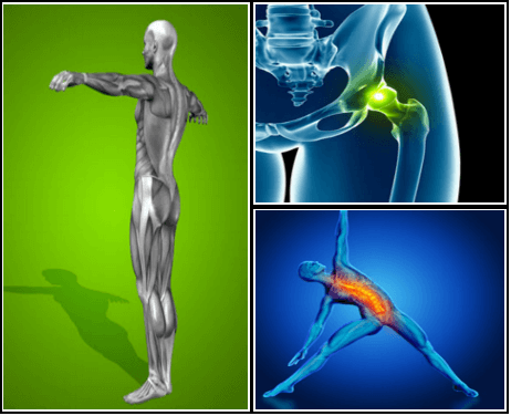
If your hips are killing you, you probably spend a lot of time sitting – in the car, at work, on that spinning seat – which puts your hips in near-constant “flexion”, says Cori Lefkowith, NASM-certified personal trainer and owner of Redefining Strength in Orange County, California. Even running involves a repetitive flexion movement that can cause pain.

Intradiscal electrothermal therapy (IDET) is a treatment for discs that are cracked or bulging as a result of degenerative disc disease. The procedure involves inserting a catheter through a small incision at the site of the disc in the back. A special wire is passed through the catheter and an electrical current is applied to heat the disc, which helps strengthen the collagen fibers of the disc wall, reducing the bulging and the related irritation of the spinal nerve. IDET is of questionable benefit.
Bursitis is inflammation of the fluid-filled sacs, called bursa, that cushion areas of pressure between joints, muscles, and tendons. Bursitis is due to overuse or repetitive actions around the joints of the body. This inflammation results in pain that is experienced during movement or pressure. Treatment involves performing stretches and strengthening exercises to help relieve pressure from the bursa.
If you have hip arthritis, work on building up the muscles in your outer thigh for added support. Lie on your pain-free side and lift the leg with arthritis up about six inches, hold for two or three seconds, and lower it again, Humphrey says. Start with one set of 10 repetitions and build up to three sets. Repeat on the other side unless it is too painful. This exercise can aggravate your symptoms if you have hip pain from bursitis.

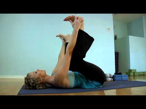
How to: Get on your hands and knees, in a tabletop position (a). Slowly widen your knees out as far as they can go and bring your feet in line with your knees. Your shins should be parallel with one another (b). Flex your feet and ease yourself forward onto your forearms. (If the stretch is too intense, try putting your arms on a block or firm pillow.) Hold for eight to 12 breaths (c). If holding the stretch for longer, try slowly moving your hips forward and backward to bring the stretch to different parts of your hips.
If all you’ve been doing for your lower body lately is the occasional lunge, it’s time to mix up your routine. “Our bodies move in different directions, not in one direction, so it’s important to mimic that during cross-training,” Whitesides says. That’s why instead of relying on a pattern of “lunge-squat-repeat” — you might want to throw some plié squats, or leg lifts into the mix.
But moving is important for hip and knee OA. It causes your joints to compress and release, bringing blood flow, nutrients, and oxygen into the cartilage. “This can help prolong the function and longevity of your joints,” says Eric Robertson, DPT, a physical therapist and associate professor of clinical physical therapy at the University of Southern California.

The diagnosis of low back pain involves a review of the history of the illness and underlying medical conditions as well as a physical examination. It is essential that a complete story of the back pain be reviewed including injury history, aggravating and alleviating conditions, associated symptoms (fever, numbness, tingling, incontinence, etc.), as well as the duration and progression of symptoms. Aside from routine abdomen and extremity evaluations, rectal and pelvic examinations may eventually be required as well. Further tests for diagnosis of low back pain can be required including blood and urine tests, plain film X-ray tests, CAT scanning, MRI scanning, bone scanning, and tests of the nerves such as electromyograms (EMG) and nerve conduction velocities (NCV).
Why is back pain still a huge problem? Maybe this: “It is extremely difficult to alter the potentially disabling belief among the lay public that low back pain has a structural mechanical cause. An important reason for this is that this belief continues to be regularly reinforced by the conditions of care of a range of ‘hands-on’ providers, for whom idiosyncratic variations of that view are fundamental to their professional existence.”

