
A.D.A.M., Inc. is accredited by URAC, also known as the American Accreditation HealthCare Commission (www.urac.org). URAC's accreditation program is an independent audit to verify that A.D.A.M. follows rigorous standards of quality and accountability. A.D.A.M. is among the first to achieve this important distinction for online health information and services. Learn more about A.D.A.M.'s editorial policy, editorial process and privacy policy. A.D.A.M. is also a founding member of Hi-Ethics and subscribes to the principles of the Health on the Net Foundation (www.hon.ch).
Before recommending exercises, physical therapists evaluate their patients to develop a routine that’s appropriate for their specific condition. Pariser says the following exercises, done at home and at the gym, are generally safe for everyone. “If a patient has already received a total hip replacement, however, certain precautions should be taken,” he says.
Avascular necrosis (also called osteonecrosis). This condition happens when blood flow to the hip bone slows and the bone tissue dies. Although it can affect other bones, avascular necrosis most often happens in the hip. It can be caused by a hip fracture or dislocation, or from the long-term use of high-dose steroids (such as prednisone), among other causes.
Spondylolisthesis. This condition occurs when one vertebra slips over the adjacent one. There are 5 types of spondylolisthesis but the most common are secondary to a defect or fracture of the pars (between the facet joints) or mechanical instability of the facet joints (degenerative). The pain can be caused by instability (back) or compression of the nerves (leg).
Long periods of inactivity in bed are no longer recommended, as this treatment may actually slow recovery. Spinal manipulation for periods of up to one month has been found to be helpful in some patients who do not have signs of nerve irritation. Future injury is avoided by using back-protection techniques during activities and support devices as needed at home or work.

Stop focusing on a specific diagnosis. Up to 85% of low back pain can be classified as "non-specific." This means that the origin of your pain cannot be localized to one specific structure or problem. While common diagnostic tests for low back pain can show the bones, discs, and joints with great detail, no test can tell the exact cause of your pain with 100% accuracy.
Magnetic resonance imaging (MRI) uses a magnetic force instead of radiation to create a computer-generated image. Unlike x-ray, which shows only bony structures, MRI scans also produce images of soft tissues such as muscles, ligaments, tendons, and blood vessels. An MRI may be ordered if a problem such as infection, tumor, inflammation, disc herniation or rupture, or pressure on a nerve is suspected. MRI is a noninvasive way to identify a condition requiring prompt surgical treatment. However, in most instances, unless there are “red flags” in the history or physical exam, an MRI scan is not necessary during the early phases of low back pain.
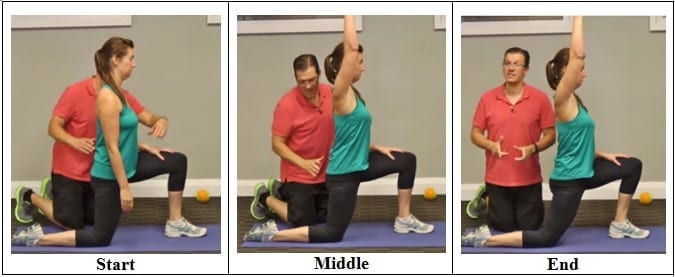
Start in a runner’s lunge, right leg forward with knee over ankle and left knee on ground with top of your foot flat on the mat. Slowly lift torso and rest hands lightly on right thigh. Lean hips forward slightly, keeping right knee behind toes, and feel the stretch in the left hip flexor. Hold here, or for a deeper stretch, raise arms overhead, biceps by ears. Hold for at least 30 seconds, then repeat on opposite side.
The hip is a basic ball-and-socket joint. The ball is the femoral head—a knob on the top of the thigh bone—and the socket is an indentation in the pelvic bone. There is cartilage lining the joint (called the labrum) and ligaments that attach the pelvic and thigh bones. Numerous muscles attach around the hip, too, moving the joint through the basic motions of flexion (bending), extension (extending the leg behind you), abduction (lifting the leg away from the body), adduction (moving the leg inward), internal rotation, and external rotation.

Radicular pain. This type of pain can occur if a spinal nerve root becomes impinged or inflamed. Radicular pain may follow a nerve root pattern or dermatome down into the buttock and/or leg. Its specific sensation is sharp, electric, burning-type pain and can be associated with numbness or weakness (sciatica). It is typically felt on only one side of the body.
I think you should mention that for some people, stretching is not the solution and that it will deteriorate their posture. Some people need stretching, but most people I know need to strengthen their "overstretched" hip flexors. Many people can't do a single hanging leg raise. Check this site if you want to know more about the importance of hip flexors bit.ly/Unlock_Your_Hip_Flexor Report
MRI (or magnetic resonance imaging) scan is a radiology technique which uses magnetism, radio waves, and a computer to produce images of body structures. MRI scanning is painless and does not involve X-ray radiation. Patients with heart pacemakers, metal implants, or metal chips or clips in or around the eyes cannot be scanned with MRI because of the effect of the magnet.
Swimming is a low impact form of exercise that can help to relieve the pressure on your joints. It’s thought that water aerobics may help to reduce the impact on your joints by up to 75%!2 Be careful though; start with a beginner’s class and always do only what you’re comfortable with – try to avoid over-exercising or pushing your muscles too far!
When was the last time you got on your gym's abductor or adductor machine and got in a good workout? It's probably been a while. Both are machines that don't get a lot of use, and they are often the target of coaches' ridicule on those "useless gym moves we should all skip" lists. Perhaps rightly so, especially if you're hopping on those machines hoping for a slimming effect.

Electrodiagnostics are procedures that, in the setting of low back pain, are primarily used to confirm whether a person has lumbar radiculopathy. The procedures include electromyography (EMG), nerve conduction studies (NCS), and evoked potential (EP) studies. EMG assesses the electrical activity in a muscle and can detect if muscle weakness results from a problem with the nerves that control the muscles. Very fine needles are inserted in muscles to measure electrical activity transmitted from the brain or spinal cord to a particular area of the body. NCSs are often performed along with EMG to exclude conditions that can mimic radiculopathy. In NCSs, two sets of electrodes are placed on the skin over the muscles. The first set provides a mild shock to stimulate the nerve that runs to a particular muscle. The second set records the nerve’s electrical signals, and from this information nerve damage that slows conduction of the nerve signal can be detected. EP tests also involve two sets of electrodes—one set to stimulate a sensory nerve, and the other placed on the scalp to record the speed of nerve signal transmissions to the brain.
Irritable hip: What you need to know Irritable hip is a common cause of hip pain and limping in children before they reach puberty. It may happen after an injury or a viral infection, or because of poor blood flow. It usually gets better with rest within 2 weeks. Pain killers may help relieve symptoms. Those under 16 years should not use aspirin. Read now
The bones of the hip are supported by specific muscles. The strongest muscle in the body is actually one of the main flexors of the hip, it is called the iliopsoas muscle. It has two attachments to the inner part of the hip as well as the lower part of the back. This muscle is involved in iliopsoas tendinitis, iliopsoas bursitis, and is one of the most common causes of muscle pain that causes hip pain. The tendons attach the muscles to the bones and the ligaments attach bone to bone. Areas that are often susceptible to increased pressure are the tendons or the muscles attached to the bone, so that is another source of pain in iliopsoas tendinitis. The bursa are pockets of fluid that also protect the top part of the bone to prevent pressure on the bone. There are about 160 bursa in the body, and when they get irritated, it can develop into bursitis.
How to do it: Grab a pair of dumbbells and stand with your feet slightly wider than hip-width apart, knees slightly bent. Hold the weights in front of your thighs, palms facing in. Maintaining a neutral spine, hinge forward from your hips, reaching the dumbbells to the ground, until your torso is almost parallel with the floor. Focus on using your glutes to raise your body halfway back up [as shown] and then return to full forward hinge again. That’s one rep. Repeat 20 times total.
A recent study from Florida Atlantic University found that doing 45 minutes of chair yoga twice per week reduced pain and improved overall quality of life in older adults with osteoarthritis. Chair yoga is practiced sitting in a chair or standing while holding the chair for support, so it’s a great option for beginners or anyone dealing with an injury or balance problems.
Health Tools Baby Due Date CalculatorBasal Metabolic Rate CalculatorBody Mass Index (BMI) CalculatorCalories Burned CalculatorChild Energy Requirements CalculatorDaily Calcium Requirements CalculatorDaily Fibre Requirements CalculatorIdeal Weight CalculatorInfectious Diseases Exclusion Periods ToolOvulation CalculatorSmoking Cost CalculatorTarget Heart Rate CalculatorWaist-to-hip Ratio Calculator Risk Tests Depression Self-AssessmentErectile Dysfunction ToolMacular Degeneration ToolOsteoporosis Risk TestProstate Symptoms Self-Assessment
The AANS does not endorse any treatments, procedures, products or physicians referenced in these patient fact sheets. This information is provided as an educational service and is not intended to serve as medical advice. Anyone seeking specific neurosurgical advice or assistance should consult his or her neurosurgeon, or locate one in your area through the AANS’ Find a Board-certified Neurosurgeon” online tool.
Prolonged sitting and activities like running or cycling can lead to tight hip flexor muscles and a variety of skeletal imbalances. Think: if you only cycle for exercise, certain muscles in your legs will get stronger (in a lot of cases you overwork these muscles) yet your core and outer hip muscles might get weaker from lack of engagement. So what? Well, these muscle imbalances often lead to skeletal imbalances and injuries down the line. If you have particularly tight hip flexors, your body will start to create an anterior pull on the pelvis (anterior pelvic tilt). You can identify an anterior pelvic tilt if your belly protrudes slightly in the front while your butt sticks out in the back (what some people refer to as “duck butt”).
Cycling: The repetitive movements of cycling can place a strain on your hip joints, not to mention that it can also affect your posture! If you really must cycle, make sure your bike is professionally fitted and that you properly warm up and stretch your hip flexors before getting on your bike. I’d recommend choosing an alternative though, or at least speaking to your doctor first!

