
Exercise appears to be useful for preventing low back pain.[47] Exercise is also probably effective in preventing recurrences in those with pain that has lasted more than six weeks.[1][48] Medium-firm mattresses are more beneficial for chronic pain than firm mattresses.[49] There is little to no evidence that back belts are any more helpful in preventing low back pain than education about proper lifting techniques.[47][50] Shoe insoles do not help prevent low back pain.[47][51]

Why is back pain still a huge problem? Maybe this: “It is extremely difficult to alter the potentially disabling belief among the lay public that low back pain has a structural mechanical cause. An important reason for this is that this belief continues to be regularly reinforced by the conditions of care of a range of ‘hands-on’ providers, for whom idiosyncratic variations of that view are fundamental to their professional existence.”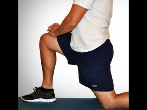
How to: Position yourself on your hands and knees, in tabletop position. Engage your abs engaged by pulling your belly button in towards your spine (a). Keeping your hips pointed towards the ground and leg bent to a 90-degree angle, raise your left knee out to the side as high as you can (b). Pause at the top, then return to starting position (c). Repeat, then switch legs.
In diagnosing the causes of hip pain, it’s important to understand hip anatomy. The hip is basically a ball-and-socket joint similar to the shoulder joint and the arms. The hip joint is impressive in that it serves two factors, it is a support structure and also very flexible. Because it is a strong support structure, it tends to get injured. Because it’s very flexible, it similarly can be susceptible to injuries.

Imaging is indicated when there are red flags, ongoing neurological symptoms that do not resolve, or ongoing or worsening pain.[5] In particular, early use of imaging (either MRI or CT) is recommended for suspected cancer, infection, or cauda equina syndrome.[5] MRI is slightly better than CT for identifying disc disease; the two technologies are equally useful for diagnosing spinal stenosis.[5] Only a few physical diagnostic tests are helpful.[5] The straight leg raise test is almost always positive in those with disc herniation.[5] Lumbar provocative discography may be useful to identify a specific disc causing pain in those with chronic high levels of low back pain.[41] Similarly, therapeutic procedures such as nerve blocks can be used to determine a specific source of pain.[5] Some evidence supports the use of facet joint injections, transforminal epidural injections and sacroilliac injections as diagnostic tests.[5] Most other physical tests, such as evaluating for scoliosis, muscle weakness or wasting, and impaired reflexes, are of little use.[5]

Traction involves the use of weights and pulleys to apply constant or intermittent force to gradually “pull” the skeletal structure into better alignment. Some people experience pain relief while in traction, but that relief is usually temporary. Once traction is released the back pain tends to return. There is no evidence that traction provides any longterm benefits for people with low back pain.
How to do it: Grab a pair of dumbbells and stand with your feet slightly wider than hip-width apart, knees slightly bent. Hold the weights in front of your thighs, palms facing in. Maintaining a neutral spine, hinge forward from your hips, reaching the dumbbells to the ground, until your torso is almost parallel with the floor. Focus on using your glutes to raise your body halfway back up [as shown] and then return to full forward hinge again. That’s one rep. Repeat 20 times total.
MRI (or magnetic resonance imaging) scan is a radiology technique which uses magnetism, radio waves, and a computer to produce images of body structures. MRI scanning is painless and does not involve X-ray radiation. Patients with heart pacemakers, metal implants, or metal chips or clips in or around the eyes cannot be scanned with MRI because of the effect of the magnet.
2016 — More editing, more! Added some better information about pain being a poor indicator, and the role of myofascial trigger points. This article has become extremely busy in the last couple months — about 4,000 readers per day, as described here — so I am really polishing it and making sure that it’s the best possible answer to people’s fears about back pain.

Too much sitting is the enemy of stiff or achy hips, says Lisa Woods, a personal trainer and yoga teacher in Eagle, Colorado. The big problem, though, isn’t just the discomfort in the sides of your thighs. It’s the chain of pain that dysfunctional hips can create, including sciatic nerve pain that can start in your lower back and go down the backs of your legs.

The information provided herein should not be used during any medical emergency or for the diagnosis or treatment of any medical condition. A licensed physician should be consulted for diagnosis and treatment of any and all medical conditions. Call 911 for all medical emergencies. Links to other sites are provided for information only -- they do not constitute endorsements of those other sites. Copyright 1997-2019, A.D.A.M., Inc. Duplication for commercial use must be authorized in writing by ADAM Health Solutions.
Nonsteroidal anti-inflammatory drugs (NSAIDS) relieve pain and inflammation and include OTC formulations (ibuprofen, ketoprofen, and naproxen sodium). Several others, including a type of NSAID called COX-2 inhibitors, are available only by prescription. Long-term use of NSAIDs has been associated with stomach irritation, ulcers, heartburn, diarrhea, fluid retention, and in rare cases, kidney dysfunction and cardiovascular disease. The longer a person uses NSAIDs the more likely they are to develop side effects. Many other drugs cannot be taken at the same time a person is treated with NSAIDs because they alter the way the body processes or eliminates other medications.
How to: Stand tall, feet slightly wider than shoulder-width apart, toes pointed out at 45-degree angles (a). Keep your back straight, knees over toes and your weight in the heels of your feet (b). Engage your glutes and thighs as you lower into a deep squat until thighs parallel to the ground (or as close as you can get them) (c). Powering through your heels, push up to return to starting position (d). Repeat.

Bone scans are used to detect and monitor infection, fracture, or disorders in the bone. A small amount of radioactive material is injected into the bloodstream and will collect in the bones, particularly in areas with some abnormality. Scanner-generated images can be used to identify specific areas of irregular bone metabolism or abnormal blood flow, as well as to measure levels of joint disease.
How to: Lie on your back with your right knee bent and foot flat on the floor (a). With your left leg fully extended, press into your right foot to shift onto your left hip. This is your starting position (b). Then, squeeze your right glutes to press your left hip open until you feel a stretch, pause, then return to start. That’s one rep (c). Perform six to eight reps, then repeat on the opposite side.

You'll need a resistance band for this one. With this exercise you're focusing on four movements—flexion, extension, abduction and adduction. Try and stand up straight while doing the exercise. If you have to lean excessively, step closer to the anchor point of your band to decrease resistance. You'll find that not only are you working the muscles of the leg that's moving, the muscles of your stance leg will work quite hard stabilizing and balancing.
MRI (or magnetic resonance imaging) scan is a radiology technique which uses magnetism, radio waves, and a computer to produce images of body structures. MRI scanning is painless and does not involve X-ray radiation. Patients with heart pacemakers, metal implants, or metal chips or clips in or around the eyes cannot be scanned with MRI because of the effect of the magnet.
I am a science writer, former massage therapist, and I was the assistant editor at ScienceBasedMedicine.org for several years. I have had my share of injuries and pain challenges as a runner and ultimate player. My wife and I live in downtown Vancouver, Canada. See my full bio and qualifications, or my blog, Writerly. You might run into me on Facebook or Twitter.
Located deep in the front of the hip and connecting the leg, pelvis, and abdomen, the hip flexors— surprise, surprise— flex the hip. But despite being some of the most powerful muscles in our bodies (with a clearly important role), it’s easy to neglect our poor hip flexors— often without even knowing it. It turns out just working at a desk all day (guilty!) can really weaken hip flexors since they tend to shorten up while in a seated position. This tightness disrupts good posture and is a common cause of lower back pain. Weakened hip flexors can also increase the risk of foot, ankle, and knee injuries (especially among runners) Hip muscle weakness and overuse injuries in recreational runners. Niemuth, P.E., Johnson, R.J., Myers, M.J., et al. Rocky Mountain University of Health Professions, Provo, VT. Clinical Journal of Sport Medicine, 2005 Jan; 15 (1): 14-21.. So be sure to get up, stand up every hour or so! And giving the hip flexors some extra attention is not just about injury prevention. Adding power to workouts, working toward greater flexibility, and getting speedier while running is also, as they say, all in the hips The effect of walking speed on muscle function and mechanical energetics. Neptune, R.R., Sasaki, K., and Kautz, S.A. Department of Mechanical Engineering, The University of Texas, Austin, TX. Gait & Posture, 2008 Jul; 28 (1): 135-43..
Epidural steroid injections are a commonly used short-term option for treating low back pain and sciatica associated with inflammation. Pain relief associated with the injections, however, tends to be temporary and the injections are not advised for long-term use. An NIH-funded randomized controlled trial assessing the benefit of epidural steroid injections for the treatment of chronic low back pain associated with spinal stenosis showed that long-term outcomes were worse among those people who received the injections compared with those who did not.
John Wolf is Onnit's Chief Fitness Officer, and an expert in unconventional training methods such as kettlebell, steel club, and suspension training. With 15-plus years of experience in the fitness industry, he has worked with rehab clients and athletes of all levels. He moves like Spider Man and can deadlift more than 500 pounds any day of the week.
Nonsteroidal anti-inflammatory drugs (NSAIDS) relieve pain and inflammation and include OTC formulations (ibuprofen, ketoprofen, and naproxen sodium). Several others, including a type of NSAID called COX-2 inhibitors, are available only by prescription. Long-term use of NSAIDs has been associated with stomach irritation, ulcers, heartburn, diarrhea, fluid retention, and in rare cases, kidney dysfunction and cardiovascular disease. The longer a person uses NSAIDs the more likely they are to develop side effects. Many other drugs cannot be taken at the same time a person is treated with NSAIDs because they alter the way the body processes or eliminates other medications.
Epidural injections of steroid drugs are frequently used to treat sciatica, despite limited evidence for their effectiveness. Moreover, these treatments are based on the assumption that reducing local inflammation in the vertebral column will relieve pain, but an association between structural abnormalities, inflammation, and sciatica symptoms has not been clearly demonstrated. NINDS-funded researchers are using a new imaging technique that can detect inflammation to better understand what causes chronic sciatica pain and to provide evidence to inform treatment selection.
Vertebroplasty and kyphoplasty are minimally invasive treatments to repair compression fractures of the vertebrae caused by osteoporosis. Vertebroplasty uses three-dimensional imaging to assist in guiding a fine needle through the skin into the vertebral body, the largest part of the vertebrae. A glue-like bone cement is then injected into the vertebral body space, which quickly hardens to stabilize and strengthen the bone and provide pain relief. In kyphoplasty, prior to injecting the bone cement, a special balloon is inserted and gently inflated to restore height to the vertebral structure and reduce spinal deformity.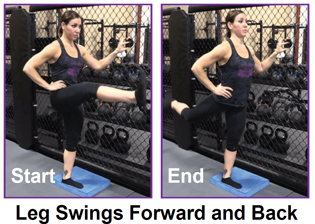
En español | You probably know someone who’s traded a worn-out hip bone for ceramic or cobalt chrome. Some 370,000 Americans undergo hip-replacement surgery each year (the average age for this is 65). But the operation isn’t a cure-all: At least 1 in 10 hip-replacement recipients will need a second procedure to repair a dislocation, mechanical failure or infection. And hip pain, with or without surgery, can be a struggle. Chronic hip pain was a factor in the accidental opioid overdoses that claimed the lives of musicians Tom Petty, 66, and Prince, 57.
The materials and information provided in this presentation, document and/or any other communication (“Communication”) from Onnit Labs, Inc. or any related entity or person (collectively “Onnit”) are strictly for informational purposes only and are not intended for use as diagnosis, prevention or treatment of a health problem or as a substitute for consulting a qualified medical professional. Some of the concepts presented herein may be theoretical.
The hip is a basic ball-and-socket joint. The ball is the femoral head—a knob on the top of the thigh bone—and the socket is an indentation in the pelvic bone. There is cartilage lining the joint (called the labrum) and ligaments that attach the pelvic and thigh bones. Numerous muscles attach around the hip, too, moving the joint through the basic motions of flexion (bending), extension (extending the leg behind you), abduction (lifting the leg away from the body), adduction (moving the leg inward), internal rotation, and external rotation.
Melissa Conrad Stöppler, MD, is a U.S. board-certified Anatomic Pathologist with subspecialty training in the fields of Experimental and Molecular Pathology. Dr. Stöppler's educational background includes a BA with Highest Distinction from the University of Virginia and an MD from the University of North Carolina. She completed residency training in Anatomic Pathology at Georgetown University followed by subspecialty fellowship training in molecular diagnostics and experimental pathology.
Back pain can be confusing enough as it is. Sometimes it may be difficult deciphering if the pain is even coming from your back, and trying to figure out what causes it can be mind-boggling. And many experts recommend focusing on mechanical sources of your back pain rather than anatomical sources of your pain. Is it a herniated disc, bulging disc, or facet joint arthritis causing your problem? It can be hard to decide, so focusing on what you are doing and the positions you put your back in may be the best way to determine the cause of your back pain.

