
Premkumar et al present evidence that the traditional “red flags” for ominous causes of back pain can be quite misleading. The correlation between red flags and ominous diagnoses is poor, and prone to producing false negatives: that is, no red flags even when there is something more serious than unexplained pain going on. In a survey of almost 10,000 patients “the absence of red flag responses did not meaningfully decrease the likelihood of a red flag diagnosis.“ This is not even remotely a surprise to anyone who paid attention in back pain school, but it’s good to have some harder data on it.
Hip fractures, or a break in the hip bone, are another common cause of hip pain. Fractures of the hip often occur after falls in the elderly patient population. Osteoporosis puts this population at increased risk for hip fractures. Stress fractures are another form of fracture that can cause hip pain. Various risk factors increase one's risk of developing a stress fracture at the hip joint.
Intervertebral disc degeneration is one of the most common mechanical causes of low back pain, and it occurs when the usually rubbery discs lose integrity as a normal process of aging. In a healthy back, intervertebral discs provide height and allow bending, flexion, and torsion of the lower back. As the discs deteriorate, they lose their cushioning ability.
If you have hip arthritis, work on building up the muscles in your outer thigh for added support. Lie on your pain-free side and lift the leg with arthritis up about six inches, hold for two or three seconds, and lower it again, Humphrey says. Start with one set of 10 repetitions and build up to three sets. Repeat on the other side unless it is too painful. This exercise can aggravate your symptoms if you have hip pain from bursitis.
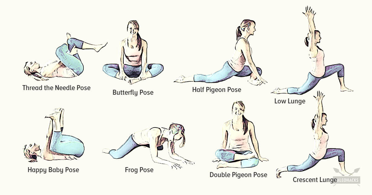
How to: Sit on the floor with knees bent so that your right shin is positioned in front of you, your left shin behind you and your left hip dropped all of the way to the floor (a). Inhale and press your left hip forward until you feel a stretch in the front of your hip (b). Exhale and press left hip back to the floor. That’s one rep (c). Complete six to eight reps, working each time to increase your range of motion. Repeat on the opposite side.
Prolonged sitting and activities like running or cycling can lead to tight hip flexor muscles and a variety of skeletal imbalances. Think: if you only cycle for exercise, certain muscles in your legs will get stronger (in a lot of cases you overwork these muscles) yet your core and outer hip muscles might get weaker from lack of engagement. So what? Well, these muscle imbalances often lead to skeletal imbalances and injuries down the line. If you have particularly tight hip flexors, your body will start to create an anterior pull on the pelvis (anterior pelvic tilt). You can identify an anterior pelvic tilt if your belly protrudes slightly in the front while your butt sticks out in the back (what some people refer to as “duck butt”).

Sit in a chair with good lumbar support and proper position and height for the task. Keep shoulders back. Switch sitting positions often and periodically walk around the office or gently stretch muscles to relieve tension. A pillow or rolled-up towel placed behind the small of the back can provide some lumbar support. During prolonged periods of sitting, elevate feet on a low stool or a stack of books.

Results after four years of follow-up showed that in general, otherwise healthy people who have surgery for one of these three conditions are likely to fare better than those who receive non-operative care. However, the results also indicated that people who are reluctant to have surgery may also recover with non-operative treatments if their conditions are not progressing and their pain is tolerable, and importantly, delaying or avoiding surgery did not cause additional damage in most cases. Researchers are continuing to track SPORT patient cohorts over a nine-year follow-up period to assess longer term treatment results and cost effectiveness across treatment options. In the interest of improving surgical techniques, NIH also is funding research on factors that contribute to the success or failure of artificial disc replacement surgery, including studies to compare discs on the market for significant differences in their durability rates over time.
Arthritis. Osteoarthritis and rheumatoid arthritis are among the most common causes of hip pain, especially in older adults. Arthritis leads to inflammation of the hip joint and the breakdown of the cartilage that cushions your hip bones. The pain gradually gets worse. People with arthritis also feel stiffness and have reduced range of motion in the hip.
The treatment of lumbar strain consists of resting the back (to avoid reinjury), medications to relieve pain and muscle spasm, local heat applications, massage, and eventual (after the acute episode resolves) reconditioning exercises to strengthen the low back and abdominal muscles. Initial treatment at home might include heat application, acetaminophen (Tylenol) or ibuprofen (Advil, Motrin), and avoiding reinjury and heavy lifting. Prescription medications that are sometimes used for acute low back pain include anti-inflammatory medications, such as sulindac (Clinoril), naproxen (Naprosyn), and ketorolac (Toradol) by injection or by mouth, muscle relaxants, such as carisoprodol (Soma), cyclobenzaprine (Flexeril), methocarbamol (Robaxin), and metaxalone (Skelaxin), as well as analgesics, such as tramadol (Ultram).
I’m not endorsing or saying these stretches are a miracle cure, but I definitely think they’ve helped. I’ve even started to add a few gentle yoga poses and pilates moves which I could so easily do, back in the day, when I was 63 kilos and participated in the greatest oxymoron ever named … a thing called the Fun Run. If I’m not careful, and I keep stretching away each day, I could end up with a 6 pack and back to my 5′ 7″ instead of 5′ 5″.

Health Tools Baby Due Date CalculatorBasal Metabolic Rate CalculatorBody Mass Index (BMI) CalculatorCalories Burned CalculatorChild Energy Requirements CalculatorDaily Calcium Requirements CalculatorDaily Fibre Requirements CalculatorIdeal Weight CalculatorInfectious Diseases Exclusion Periods ToolOvulation CalculatorSmoking Cost CalculatorTarget Heart Rate CalculatorWaist-to-hip Ratio Calculator Risk Tests Depression Self-AssessmentErectile Dysfunction ToolMacular Degeneration ToolOsteoporosis Risk TestProstate Symptoms Self-Assessment
Nerve block therapies aim to relieve chronic pain by blocking nerve conduction from specific areas of the body. Nerve block approaches range from injections of local anesthetics, botulinum toxin, or steroids into affected soft tissues or joints to more complex nerve root blocks and spinal cord stimulation. When extreme pain is involved, low doses of drugs may be administered by catheter directly into the spinal cord. The success of a nerve block approach depends on the ability of a practitioner to locate and inject precisely the correct nerve. Chronic use of steroid injections may lead to increased functional impairment. 
MRI and x-ray for low back pain are surprisingly unreliable,1 because things like bulging discs usually aren’t a deal,2 most back pain goes away on its own,3 and trigger points (“muscle knots”) are common and can be alarmingly intense but aren’t dangerous.4 Most patients are much better off when they feel confident about these things. The power of justified, rational confidence is a huge factor in back pain.5 Sadly, many healthcare professionals continue to perpetuate the idea of fragile backs,6 which undermines that valuable confidence.
Injury to the bones and joints: Fractures (breakage of bone) of the lumbar spine and sacrum bone most commonly affect elderly people with osteoporosis, especially those who have taken long-term cortisone medication. For these individuals, occasionally even minimal stresses on the spine (such as bending to tie shoes) can lead to bone fracture. In this setting, the vertebra can collapse (vertebral compression fracture). The fracture causes an immediate onset of severe localized pain that can radiate around the waist in a band-like fashion and is made intensely worse with body motions. This pain generally does not radiate down the lower extremities. Vertebral fractures in younger patients occur only after severe trauma, such as from motor-vehicle accidents or a convulsive seizure.
Lay on your back on your mat and pull your knees to your chest. Place your hands on the inside arches of your feet and open your knees wider than shoulder-width apart. Keeping your back pressed into the mat as much as possible, press your feet into hands while pulling down on feet, creating resistance. Breathe deeply and hold for at least 30 seconds.
In diagnosing the causes of hip pain, it’s important to understand hip anatomy. The hip is basically a ball-and-socket joint similar to the shoulder joint and the arms. The hip joint is impressive in that it serves two factors, it is a support structure and also very flexible. Because it is a strong support structure, it tends to get injured. Because it’s very flexible, it similarly can be susceptible to injuries.

To understand various causes of low back pain, it is important to appreciate the normal design (anatomy) of the tissues of this area of the body. Important structures of the low back that can be related to symptoms in this region include the bony lumbar spine (vertebrae, singular = vertebra), discs between the vertebrae, ligaments around the spine and discs, spinal cord and nerves, muscles of the low back, internal organs of the pelvis and abdomen, and the skin covering the lumbar area.
Antidepressants may be effective for treating chronic pain associated with symptoms of depression, but they have a risk of side effects.[13] Although the antiseizure drugs gabapentin, pregabalin, and topiramate are sometimes used for chronic low back pain evidence does not support a benefit.[79] Systemic oral steroids have not been shown to be useful in low back pain.[1][13] Facet joint injections and steroid injections into the discs have not been found to be effective in those with persistent, non-radiating pain; however, they may be considered for those with persistent sciatic pain.[80] Epidural corticosteroid injections provide a slight and questionable short-term improvement in those with sciatica but are of no long term benefit.[81] There are also concerns of potential side effects.[82]
Chronic back pain is defined as pain that persists for 12 weeks or longer, even after an initial injury or underlying cause of acute low back pain has been treated. About 20 percent of people affected by acute low back pain develop chronic low back pain with persistent symptoms at one year. In some cases, treatment successfully relieves chronic low back pain, but in other cases pain persists despite medical and surgical treatment.
Stretching the hip muscles that sit on top of the bursae, part of the lining in your hip joint, can give you some relief from bursitis pain. Kneel on the leg that's giving you the pain, holding on to something sturdy for balance. Tilt your pelvis forward, tightening your gluteus muscles (the muscles in your buttocks). Then lean away from the side of your hip that hurts, for instance to the left if you're kneeling on your right knee. You should feel a stretch from the top of your hip bone down the side of your leg to your knee, Humphrey says. Hold the stretch for 30 seconds and repeat once or twice.
Approximately 15 degrees of hip extension is required to walk normally. If hip flexors are tight then in order to walk, compensatory movement needs to take place through the lower back causing back pain and premature disc degeneration. Like other joints, if we fail to take them through their full range on a regular basis we eventually lose mobility.
Health Tools Baby Due Date CalculatorBasal Metabolic Rate CalculatorBody Mass Index (BMI) CalculatorCalories Burned CalculatorChild Energy Requirements CalculatorDaily Calcium Requirements CalculatorDaily Fibre Requirements CalculatorIdeal Weight CalculatorInfectious Diseases Exclusion Periods ToolOvulation CalculatorSmoking Cost CalculatorTarget Heart Rate CalculatorWaist-to-hip Ratio Calculator Risk Tests Depression Self-AssessmentErectile Dysfunction ToolMacular Degeneration ToolOsteoporosis Risk TestProstate Symptoms Self-Assessment
Imaging is indicated when there are red flags, ongoing neurological symptoms that do not resolve, or ongoing or worsening pain.[5] In particular, early use of imaging (either MRI or CT) is recommended for suspected cancer, infection, or cauda equina syndrome.[5] MRI is slightly better than CT for identifying disc disease; the two technologies are equally useful for diagnosing spinal stenosis.[5] Only a few physical diagnostic tests are helpful.[5] The straight leg raise test is almost always positive in those with disc herniation.[5] Lumbar provocative discography may be useful to identify a specific disc causing pain in those with chronic high levels of low back pain.[41] Similarly, therapeutic procedures such as nerve blocks can be used to determine a specific source of pain.[5] Some evidence supports the use of facet joint injections, transforminal epidural injections and sacroilliac injections as diagnostic tests.[5] Most other physical tests, such as evaluating for scoliosis, muscle weakness or wasting, and impaired reflexes, are of little use.[5]
Exercise appears to be useful for preventing low back pain.[47] Exercise is also probably effective in preventing recurrences in those with pain that has lasted more than six weeks.[1][48] Medium-firm mattresses are more beneficial for chronic pain than firm mattresses.[49] There is little to no evidence that back belts are any more helpful in preventing low back pain than education about proper lifting techniques.[47][50] Shoe insoles do not help prevent low back pain.[47][51]
Lumbar radiculopathy: Lumbar radiculopathy is nerve irritation that is caused by damage to the discs between the vertebrae. Damage to the disc occurs because of degeneration ("wear and tear") of the outer ring of the disc, traumatic injury, or both. As a result, the central softer portion of the disc can rupture (herniate) through the outer ring of the disc and abut the spinal cord or its nerves as they exit the bony spinal column. This rupture is what causes the commonly recognized "sciatica" pain of a herniated disc that shoots from the low back and buttock down the leg. Sciatica can be preceded by a history of localized low-back aching or it can follow a "popping" sensation and be accompanied by numbness and tingling. The pain commonly increases with movements at the waist and can increase with coughing or sneezing. In more severe instances, sciatica can be accompanied by incontinence of the bladder and/or bowels. The sciatica of lumbar radiculopathy typically affects only one side of the body, such as the left side or right side, and not both. Lumbar radiculopathy is suspected based on the above symptoms. Increased radiating pain when the lower extremity is lifted supports the diagnosis. Nerve testing (EMG/electromyogramspina bifida
Low back pain may be classified based on the signs and symptoms. Diffuse pain that does not change in response to particular movements, and is localized to the lower back without radiating beyond the buttocks, is classified as nonspecific, the most common classification.[5] Pain that radiates down the leg below the knee, is located on one side (in the case of disc herniation), or is on both sides (in spinal stenosis), and changes in severity in response to certain positions or maneuvers is radicular, making up 7% of cases.[5] Pain that is accompanied by red flags such as trauma, fever, a history of cancer or significant muscle weakness may indicate a more serious underlying problem and is classified as needing urgent or specialized attention.[5]
Emerging technologies such as X-rays gave physicians new diagnostic tools, revealing the intervertebral disc as a source for back pain in some cases. In 1938, orthopedic surgeon Joseph S. Barr reported on cases of disc-related sciatica improved or cured with back surgery.[100] As a result of this work, in the 1940s, the vertebral disc model of low back pain took over,[99] dominating the literature through the 1980s, aiding further by the rise of new imaging technologies such as CT and MRI.[100] The discussion subsided as research showed disc problems to be a relatively uncommon cause of the pain. Since then, physicians have come to realize that it is unlikely that a specific cause for low back pain can be identified in many cases and question the need to find one at all as most of the time symptoms resolve within 6 to 12 weeks regardless of treatment.[99]
You could do these moves all together as a single workout, or, as Miranda suggests, split them in half and do the first part one day and the second part another—"but do the warm-up with each one," she says. Those first three moves are meant to not only "wake up" the muscles, but also get your brain ready for the movement patterns to come. For that reason, she says that doing the first three moves "would be a fantastic warm-up before any workout."

